 Movie
Movie Controller

[English] 日本語
 Yorodumi
- PDB-6s6h: Crystal structure of the DNA binding domain of the chromosome-par... -
+ Open data

- Basic information
| Entry | Database: PDB / ID: 6s6h | ||||||||||||||||||
|---|---|---|---|---|---|---|---|---|---|---|---|---|---|---|---|---|---|---|---|
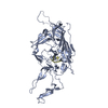
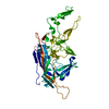
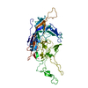
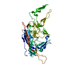
| Title | Crystal structure of the DNA binding domain of the chromosome-partitioning protein ParB complexed to the centromeric parS site | ||||||||||||||||||
 Components Components |
| ||||||||||||||||||
 Keywords Keywords | DNA BINDING PROTEIN / chromosome segregation / chromosome maintenance / protein-DNA recognition / evolution / DNA-binding protein | ||||||||||||||||||
| Function / homology |  Function and homology information Function and homology informationpositive regulation of sporulation resulting in formation of a cellular spore / chromosome segregation / chromosome / DNA binding / cytoplasm Similarity search - Function | ||||||||||||||||||
| Biological species |  Caulobacter vibrioides NA1000 (bacteria)Caulobacter vibrioides (bacteria) Caulobacter vibrioides NA1000 (bacteria)Caulobacter vibrioides (bacteria) | ||||||||||||||||||
| Method |  X-RAY DIFFRACTION / SYNCHROTRON / MOLECULAR REPLACEMENT / Resolution: 2.4 Å X-RAY DIFFRACTION / SYNCHROTRON / MOLECULAR REPLACEMENT / Resolution: 2.4 Å | ||||||||||||||||||
 Authors Authors | Jalal, A.S.B. / Tran, N.T. / Stevenson, C.E.M. / Tan, E.X. / Lawson, D.M. / Le, T.B.K. | ||||||||||||||||||
| Funding support |  United Kingdom, 5items United Kingdom, 5items
| ||||||||||||||||||
 Citation Citation | Journal: Cell Rep / Year: 2020 Title: Diversification of DNA-Binding Specificity by Permissive and Specificity-Switching Mutations in the ParB/Noc Protein Family. Authors: Jalal, A.S.B. / Tran, N.T. / Stevenson, C.E. / Chan, E.W. / Lo, R. / Tan, X. / Noy, A. / Lawson, D.M. / Le, T.B.K. | ||||||||||||||||||
| History |
|
- Structure visualization
| Structure viewer | Molecule: MolmilJmol/JSmol |
|---|
- Downloads & links
-Download
| PDBx/mmCIF format | 6s6h.cif.gz | 155.7 KB | Display | PDBx/mmCIF format |
|---|---|---|---|---|
| PDB format | pdb6s6h.ent.gz | 118.6 KB | Display | PDB format |
| PDBx/mmJSON format | 6s6h.json.gz | Tree view | PDBx/mmJSON format | |
| Others |  Other downloads Other downloads |
-Validation report
| Arichive directory | https://data.pdbj.org/pub/pdb/validation_reports/s6/6s6hftp://data.pdbj.org/pub/pdb/validation_reports/s6/6s6h | HTTPS FTP |
|---|
-Related structure data
| Related structure data | 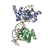 6y93C 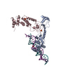 4umkS S: Starting model for refinement C: citing same article ( |
|---|---|
| Similar structure data |


-Links
 PDBj
PDBj


- Assembly
| Deposited unit | 
| ||||||||||||||||||||||||||||||||||||||||||||||||||||||||||||||||||||
|---|---|---|---|---|---|---|---|---|---|---|---|---|---|---|---|---|---|---|---|---|---|---|---|---|---|---|---|---|---|---|---|---|---|---|---|---|---|---|---|---|---|---|---|---|---|---|---|---|---|---|---|---|---|---|---|---|---|---|---|---|---|---|---|---|---|---|---|---|---|
| 1 |
| ||||||||||||||||||||||||||||||||||||||||||||||||||||||||||||||||||||
| Unit cell |
| ||||||||||||||||||||||||||||||||||||||||||||||||||||||||||||||||||||
| Noncrystallographic symmetry (NCS) | NCS domain:
NCS domain segments: Component-ID: _ / Refine code: _
NCS ensembles :
|
-Components
| #1: Protein | Mass: 15502.797 Da / Num. of mol.: 2 Source method: isolated from a genetically manipulated source Details: The expressed protein comprised residues 126-254 of the wild-type sequence with an additional N-terminal Met residue and a C-terminal nickel affinity tag of sequence KLAAALEHHHHHH from the ...Details: The expressed protein comprised residues 126-254 of the wild-type sequence with an additional N-terminal Met residue and a C-terminal nickel affinity tag of sequence KLAAALEHHHHHH from the pET21b expression plasmid. Source: (gene. exp.) Caulobacter vibrioides NA1000 (bacteria)Gene: parB, CCNA_03868 / Plasmid: pET21b / Production host: #2: DNA chain | Mass: 6132.991 Da / Num. of mol.: 2 / Source method: obtained synthetically Details: Chains C and D form a symmetrical DNA duplex which is similar to the parS sequence from Caulobacter vibrioides without 5' phosphates Source: (synth.) Caulobacter vibrioides (bacteria)#3: Chemical |   Mass: 92.094 Da / Num. of mol.: 2 / Source method: obtained synthetically / Formula: C3H8O3 Mass: 92.094 Da / Num. of mol.: 2 / Source method: obtained synthetically / Formula: C3H8O3#4: Water | ChemComp-HOH / |  Mass: 18.015 Da / Num. of mol.: 82 / Source method: isolated from a natural source / Formula: H2O Mass: 18.015 Da / Num. of mol.: 82 / Source method: isolated from a natural source / Formula: H2OHas ligand of interest | N | |
|---|
-Experimental details
-Experiment
| Experiment | Method: X-RAY DIFFRACTION / Number of used crystals: 1 |
|---|
- Sample preparation
| Crystal | Density Matthews: 2.4 Å3/Da / Density % sol: 48.73 % |
|---|---|
| Crystal grow | Temperature: 293 K / Method: vapor diffusion, hanging drop / pH: 8 / Details: NULL |
-Data collection
| Diffraction | Mean temperature: 100 K / Serial crystal experiment: N | |||||||||||||||||||||||||||
|---|---|---|---|---|---|---|---|---|---|---|---|---|---|---|---|---|---|---|---|---|---|---|---|---|---|---|---|---|
| Diffraction source | Source: SYNCHROTRON / Site: Diamond / Beamline: I04 / Wavelength: 0.9795 Å | |||||||||||||||||||||||||||
| Detector | Type: DECTRIS PILATUS 6M-F / Detector: PIXEL / Date: Oct 24, 2018 | |||||||||||||||||||||||||||
| Radiation | Protocol: SINGLE WAVELENGTH / Monochromatic (M) / Laue (L): M / Scattering type: x-ray | |||||||||||||||||||||||||||
| Radiation wavelength | Wavelength: 0.9795 Å / Relative weight: 1 | |||||||||||||||||||||||||||
| Reflection | Resolution: 2.4→40.12 Å / Num. obs: 16317 / % possible obs: 99.7 % / Redundancy: 6.4 % / CC1/2: 0.992 / Rmerge(I) obs: 0.137 / Rpim(I) all: 0.058 / Rrim(I) all: 0.15 / Net I/σ(I): 7 / Num. measured all: 105021 / Scaling rejects: 34 | |||||||||||||||||||||||||||
| Reflection shell | Diffraction-ID: 1 / % possible all: 99.2
|
- Processing
| Software |
| |||||||||||||||||||||||||||||||||||||||||||||||||||||||||||||||||||||||||||||||||||||||||||||||||||||||||||||||||||||||||||||
|---|---|---|---|---|---|---|---|---|---|---|---|---|---|---|---|---|---|---|---|---|---|---|---|---|---|---|---|---|---|---|---|---|---|---|---|---|---|---|---|---|---|---|---|---|---|---|---|---|---|---|---|---|---|---|---|---|---|---|---|---|---|---|---|---|---|---|---|---|---|---|---|---|---|---|---|---|---|---|---|---|---|---|---|---|---|---|---|---|---|---|---|---|---|---|---|---|---|---|---|---|---|---|---|---|---|---|---|---|---|---|---|---|---|---|---|---|---|---|---|---|---|---|---|---|---|---|
| Refinement | Method to determine structure: MOLECULAR REPLACEMENT Starting model: 4UMK Resolution: 2.4→40.12 Å / Cor.coef. Fo:Fc: 0.944 / Cor.coef. Fo:Fc free: 0.942 / SU B: 19.018 / SU ML: 0.21 / SU R Cruickshank DPI: 0.4347 / Cross valid method: THROUGHOUT / σ(F): 0 / ESU R: 0.435 / ESU R Free: 0.243 Details: HYDROGENS HAVE BEEN ADDED IN THE RIDING POSITIONS U VALUES : WITH TLS ADDED
| |||||||||||||||||||||||||||||||||||||||||||||||||||||||||||||||||||||||||||||||||||||||||||||||||||||||||||||||||||||||||||||
| Solvent computation | Ion probe radii: 0.8 Å / Shrinkage radii: 0.8 Å / VDW probe radii: 1.2 Å | |||||||||||||||||||||||||||||||||||||||||||||||||||||||||||||||||||||||||||||||||||||||||||||||||||||||||||||||||||||||||||||
| Displacement parameters | Biso max: 135.81 Å2 / Biso mean: 48.259 Å2 / Biso min: 23.22 Å2
| |||||||||||||||||||||||||||||||||||||||||||||||||||||||||||||||||||||||||||||||||||||||||||||||||||||||||||||||||||||||||||||
| Refinement step | Cycle: final / Resolution: 2.4→40.12 Å
| |||||||||||||||||||||||||||||||||||||||||||||||||||||||||||||||||||||||||||||||||||||||||||||||||||||||||||||||||||||||||||||
| Refine LS restraints |
| |||||||||||||||||||||||||||||||||||||||||||||||||||||||||||||||||||||||||||||||||||||||||||||||||||||||||||||||||||||||||||||
| Refine LS restraints NCS | Refine-ID: X-RAY DIFFRACTION / Type: interatomic distance / Weight position: 0.05
| |||||||||||||||||||||||||||||||||||||||||||||||||||||||||||||||||||||||||||||||||||||||||||||||||||||||||||||||||||||||||||||
| LS refinement shell | Resolution: 2.4→2.462 Å / Rfactor Rfree error: 0 / Total num. of bins used: 20
| |||||||||||||||||||||||||||||||||||||||||||||||||||||||||||||||||||||||||||||||||||||||||||||||||||||||||||||||||||||||||||||
| Refinement TLS params. | Method: refined / Refine-ID: X-RAY DIFFRACTION
| |||||||||||||||||||||||||||||||||||||||||||||||||||||||||||||||||||||||||||||||||||||||||||||||||||||||||||||||||||||||||||||
| Refinement TLS group |
|
