 Movie
Movie Controller

[English] 日本語
 Yorodumi
- PDB-6s3x: Crystal Structure of the Homospermidine Synthase (HSS) variant E2... -
+ Open data

- Basic information
| Entry | Database: PDB / ID: 6s3x | |||||||||
|---|---|---|---|---|---|---|---|---|---|---|
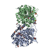
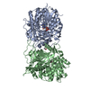
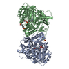
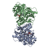
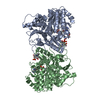
| Title | Crystal Structure of the Homospermidine Synthase (HSS) variant E210Q from Blastochloris viridis in Complex with NAD | |||||||||
 Components Components | Homospermidine synthase | |||||||||
 Keywords Keywords | TRANSFERASE / Homospermidine Synthase / Rossmann Fold / NAD / Putrescine | |||||||||
| Function / homology |  Function and homology information Function and homology informationhomospermidine synthase (spermidine-specific) activity / homospermidine synthase / homospermidine synthase activity Similarity search - Function | |||||||||
| Biological species |  Blastochloris viridis (bacteria) Blastochloris viridis (bacteria) | |||||||||
| Method |  X-RAY DIFFRACTION / SYNCHROTRON / MOLECULAR REPLACEMENT / Resolution: 1.72 Å X-RAY DIFFRACTION / SYNCHROTRON / MOLECULAR REPLACEMENT / Resolution: 1.72 Å | |||||||||
 Authors Authors | Helfrich, F. / Scheidig, A.J. | |||||||||
 Citation Citation | Journal: Acta Crystallogr D Struct Biol / Year: 2021 Title: Structural and catalytic characterization of Blastochloris viridis and Pseudomonas aeruginosa homospermidine synthases supports the essential role of cation-pi interaction. Authors: Helfrich, F. / Scheidig, A.J. | |||||||||
| History |
|
- Structure visualization
| Structure viewer | Molecule: MolmilJmol/JSmol |
|---|
- Downloads & links
-Download
| PDBx/mmCIF format | 6s3x.cif.gz | 376.5 KB | Display | PDBx/mmCIF format |
|---|---|---|---|---|
| PDB format | pdb6s3x.ent.gz | 308.8 KB | Display | PDB format |
| PDBx/mmJSON format | 6s3x.json.gz | Tree view | PDBx/mmJSON format | |
| Others |  Other downloads Other downloads |
-Validation report
| Arichive directory | https://data.pdbj.org/pub/pdb/validation_reports/s3/6s3xftp://data.pdbj.org/pub/pdb/validation_reports/s3/6s3x | HTTPS FTP |
|---|
-Related structure data
| Related structure data | 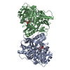 6s49C  6s4dC 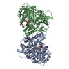 6s6gC  6s72C  6sepC 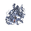 6y87C  4plpS S: Starting model for refinement C: citing same article ( |
|---|---|
| Similar structure data |

-Links
 PDBj
PDBj- Assembly
| Deposited unit | 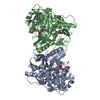
| ||||||||
|---|---|---|---|---|---|---|---|---|---|
| 1 |
| ||||||||
| Unit cell |
|
-Components
| #1: Protein | Mass: 53036.508 Da / Num. of mol.: 2 / Mutation: E210Q Source method: isolated from a genetically manipulated source Source: (gene. exp.) Blastochloris viridis (bacteria) / Gene: hss / Production host: #2: Chemical |   Mass: 96.063 Da / Num. of mol.: 2 / Source method: isolated from a natural source / Formula: SO4 Mass: 96.063 Da / Num. of mol.: 2 / Source method: isolated from a natural source / Formula: SO4#3: Chemical |   Mass: 130.191 Da / Num. of mol.: 2 / Source method: isolated from a natural source / Formula: C5H14N4 Mass: 130.191 Da / Num. of mol.: 2 / Source method: isolated from a natural source / Formula: C5H14N4#4: Chemical |   Mass: 663.425 Da / Num. of mol.: 2 / Source method: isolated from a natural source / Formula: C21H27N7O14P2 / Feature type: SUBJECT OF INVESTIGATION / Comment: NAD*YM Mass: 663.425 Da / Num. of mol.: 2 / Source method: isolated from a natural source / Formula: C21H27N7O14P2 / Feature type: SUBJECT OF INVESTIGATION / Comment: NAD*YM#5: Water | ChemComp-HOH / |  Mass: 18.015 Da / Num. of mol.: 846 / Source method: isolated from a natural source / Formula: H2O Mass: 18.015 Da / Num. of mol.: 846 / Source method: isolated from a natural source / Formula: H2OHas ligand of interest | Y | |
|---|
-Experimental details
-Experiment
| Experiment | Method: X-RAY DIFFRACTION / Number of used crystals: 1 |
|---|
- Sample preparation
| Crystal | Density Matthews: 2.5 Å3/Da / Density % sol: 50.84 % |
|---|---|
| Crystal grow | Temperature: 291 K / Method: vapor diffusion, hanging drop / pH: 4.8 Details: Sodium acetate, Ammonium acetate, PEG 10000, NDSB-201, Agmatine sulfate |
-Data collection
| Diffraction | Mean temperature: 100 K / Serial crystal experiment: N |
|---|---|
| Diffraction source | Source: SYNCHROTRON / Site: PETRA III, EMBL c/o DESY  / Beamline: P14 (MX2) / Wavelength: 0.9789 Å / Beamline: P14 (MX2) / Wavelength: 0.9789 Å |
| Detector | Type: DECTRIS EIGER X 16M / Detector: PIXEL / Date: Oct 10, 2017 |
| Radiation | Protocol: SINGLE WAVELENGTH / Monochromatic (M) / Laue (L): M / Scattering type: x-ray |
| Radiation wavelength | Wavelength: 0.9789 Å / Relative weight: 1 |
| Reflection | Resolution: 1.66→90.36 Å / Num. obs: 111532 / % possible obs: 89.5 % / Redundancy: 7 % / Rrim(I) all: 0.197 / Net I/σ(I): 6.4 |
| Reflection shell | Resolution: 1.66→1.69 Å / Num. unique obs: 1004 |
- Processing
| Software |
| |||||||||||||||||||||||||||||||||||||||||||||||||||||||||||||||||||||||||||||||||||||||||||||||||||||||||||||||||||||||||||||||||||||||||||||||||||||||||||||||||||||||||||||||||||||||||||||||||||||||||||||||||||||||||
|---|---|---|---|---|---|---|---|---|---|---|---|---|---|---|---|---|---|---|---|---|---|---|---|---|---|---|---|---|---|---|---|---|---|---|---|---|---|---|---|---|---|---|---|---|---|---|---|---|---|---|---|---|---|---|---|---|---|---|---|---|---|---|---|---|---|---|---|---|---|---|---|---|---|---|---|---|---|---|---|---|---|---|---|---|---|---|---|---|---|---|---|---|---|---|---|---|---|---|---|---|---|---|---|---|---|---|---|---|---|---|---|---|---|---|---|---|---|---|---|---|---|---|---|---|---|---|---|---|---|---|---|---|---|---|---|---|---|---|---|---|---|---|---|---|---|---|---|---|---|---|---|---|---|---|---|---|---|---|---|---|---|---|---|---|---|---|---|---|---|---|---|---|---|---|---|---|---|---|---|---|---|---|---|---|---|---|---|---|---|---|---|---|---|---|---|---|---|---|---|---|---|---|---|---|---|---|---|---|---|---|---|---|---|---|---|---|---|---|
| Refinement | Method to determine structure: MOLECULAR REPLACEMENT Starting model: 4PLP Resolution: 1.72→90.36 Å / SU ML: 0.2 / Cross valid method: FREE R-VALUE / σ(F): 1.34 / Phase error: 21.45
| |||||||||||||||||||||||||||||||||||||||||||||||||||||||||||||||||||||||||||||||||||||||||||||||||||||||||||||||||||||||||||||||||||||||||||||||||||||||||||||||||||||||||||||||||||||||||||||||||||||||||||||||||||||||||
| Solvent computation | Shrinkage radii: 0.9 Å / VDW probe radii: 1.11 Å | |||||||||||||||||||||||||||||||||||||||||||||||||||||||||||||||||||||||||||||||||||||||||||||||||||||||||||||||||||||||||||||||||||||||||||||||||||||||||||||||||||||||||||||||||||||||||||||||||||||||||||||||||||||||||
| Refinement step | Cycle: LAST / Resolution: 1.72→90.36 Å
| |||||||||||||||||||||||||||||||||||||||||||||||||||||||||||||||||||||||||||||||||||||||||||||||||||||||||||||||||||||||||||||||||||||||||||||||||||||||||||||||||||||||||||||||||||||||||||||||||||||||||||||||||||||||||
| Refine LS restraints |
| |||||||||||||||||||||||||||||||||||||||||||||||||||||||||||||||||||||||||||||||||||||||||||||||||||||||||||||||||||||||||||||||||||||||||||||||||||||||||||||||||||||||||||||||||||||||||||||||||||||||||||||||||||||||||
| LS refinement shell |
|
