 Movie
Movie Controller

[English] 日本語
 Yorodumi
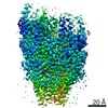
- PDB-6s3r: Structure of the FliPQR complex from the flagellar type 3 secreti... -
+ Open data

- Basic information
| Entry | Database: PDB / ID: 6s3r | ||||||||||||||||||
|---|---|---|---|---|---|---|---|---|---|---|---|---|---|---|---|---|---|---|---|
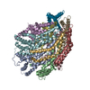
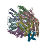
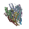
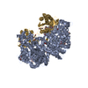
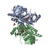
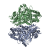
| Title | Structure of the FliPQR complex from the flagellar type 3 secretion system of Pseudomonas savastanoi. | ||||||||||||||||||
 Components Components |
| ||||||||||||||||||
 Keywords Keywords | PROTEIN TRANSPORT / flagella / T3SS / export apparatus / export gate | ||||||||||||||||||
| Function / homology |  Function and homology information Function and homology informationbacterial-type flagellum organization / bacterial-type flagellum basal body / bacterial-type flagellum assembly / protein secretion / protein targeting / plasma membrane Similarity search - Function | ||||||||||||||||||
| Biological species |  Pseudomonas savastanoi pv. phaseolicola (bacteria) Pseudomonas savastanoi pv. phaseolicola (bacteria) | ||||||||||||||||||
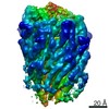
| Method | ELECTRON MICROSCOPY / single particle reconstruction / cryo EM / Resolution: 3.5 Å | ||||||||||||||||||
 Authors Authors | Kuhlen, L. / Johnson, S. / Deme, J.C. / Lea, S.M. | ||||||||||||||||||
| Funding support |  United Kingdom, 5items United Kingdom, 5items
| ||||||||||||||||||
 Citation Citation | Journal: Nat Commun / Year: 2020 Title: The substrate specificity switch FlhB assembles onto the export gate to regulate type three secretion. Authors: Lucas Kuhlen / Steven Johnson / Andreas Zeitler / Sandra Bäurle / Justin C Deme / Joseph J E Caesar / Rebecca Debo / Joseph Fisher / Samuel Wagner / Susan M Lea /  Abstract: Protein secretion through type-three secretion systems (T3SS) is critical for motility and virulence of many bacteria. Proteins are transported through an export gate containing three proteins ...Protein secretion through type-three secretion systems (T3SS) is critical for motility and virulence of many bacteria. Proteins are transported through an export gate containing three proteins (FliPQR in flagella, SctRST in virulence systems). A fourth essential T3SS protein (FlhB/SctU) functions to "switch" secretion substrate specificity once the growing hook/needle reach their determined length. Here, we present the cryo-electron microscopy structure of an export gate containing the switch protein from a Vibrio flagellar system at 3.2 Å resolution. The structure reveals that FlhB/SctU extends the helical export gate with its four predicted transmembrane helices wrapped around FliPQR/SctRST. The unusual topology of the FlhB/SctU helices creates a loop wrapped around the bottom of the closed export gate. Structure-informed mutagenesis suggests that this loop is critical in gating secretion and we propose that a series of conformational changes in the T3SS trigger opening of the gate through interactions between FlhB/SctU and FliPQR/SctRST. | ||||||||||||||||||
| History |
|
- Structure visualization
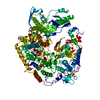
| Movie |
Movie viewer |
|---|---|
| Structure viewer | Molecule: MolmilJmol/JSmol |
- Downloads & links
-Download
| PDBx/mmCIF format | 6s3r.cif.gz | 293 KB | Display | PDBx/mmCIF format |
|---|---|---|---|---|
| PDB format | pdb6s3r.ent.gz | 232.8 KB | Display | PDB format |
| PDBx/mmJSON format | 6s3r.json.gz | Tree view | PDBx/mmJSON format | |
| Others |  Other downloads Other downloads |
-Validation report
| Arichive directory | https://data.pdbj.org/pub/pdb/validation_reports/s3/6s3rftp://data.pdbj.org/pub/pdb/validation_reports/s3/6s3r | HTTPS FTP |
|---|
-Related structure data
| Related structure data | 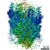 10095MC 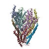 6s3lC 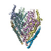 6s3sC M: map data used to model this data C: citing same article ( |
|---|---|
| Similar structure data |

-Links
 PDBj
PDBj
- Assembly
| Deposited unit | 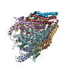
|
|---|---|
| 1 |
|
-Components
| #1: Protein | Mass: 27199.805 Da / Num. of mol.: 5 Source method: isolated from a genetically manipulated source Source: (gene. exp.) Pseudomonas savastanoi pv. phaseolicola (strain 1448A / Race 6) (bacteria)Strain: 1448A / Race 6 / Gene: fliP, PSPPH_3370 / Production host: #2: Protein | | Mass: 32352.314 Da / Num. of mol.: 1 Source method: isolated from a genetically manipulated source Source: (gene. exp.) Pseudomonas savastanoi pv. phaseolicola (bacteria)Gene: ALQ02_02028 / Production host: #3: Protein | Mass: 9925.104 Da / Num. of mol.: 5 Source method: isolated from a genetically manipulated source Source: (gene. exp.) Pseudomonas savastanoi pv. phaseolicola (strain 1448A / Race 6) (bacteria)Strain: 1448A / Race 6 / Gene: fliQ, PSPPH_3369 / Production host: |
|---|
-Experimental details
-Experiment
| Experiment | Method: ELECTRON MICROSCOPY |
|---|---|
| EM experiment | Aggregation state: PARTICLE / 3D reconstruction method: single particle reconstruction |
- Sample preparation
| Component | Name: FliPQR complex from the flagellar type 3 secretion system of Pseudomonas savastanoi Type: COMPLEX / Entity ID: all / Source: RECOMBINANT | |||||||||||||||||||||||||
|---|---|---|---|---|---|---|---|---|---|---|---|---|---|---|---|---|---|---|---|---|---|---|---|---|---|---|
| Molecular weight | Value: 0.21 MDa / Experimental value: YES | |||||||||||||||||||||||||
| Source (natural) | Organism: Pseudomonas savastanoi pv. phaseolicola 1448A (bacteria) | |||||||||||||||||||||||||
| Source (recombinant) | Organism: | |||||||||||||||||||||||||
| Buffer solution | pH: 8 | |||||||||||||||||||||||||
| Buffer component |
| |||||||||||||||||||||||||
| Specimen | Conc.: 4.1 mg/ml / Embedding applied: NO / Shadowing applied: NO / Staining applied: NO / Vitrification applied: YES | |||||||||||||||||||||||||
| Specimen support | Grid material: GOLD / Grid mesh size: 300 divisions/in. / Grid type: Quantifoil R1.2/1.3 | |||||||||||||||||||||||||
| Vitrification | Instrument: FEI VITROBOT MARK IV / Cryogen name: ETHANE / Humidity: 100 % / Chamber temperature: 295 K / Details: 10 seconds wait time before blotting |
- Electron microscopy imaging
| Experimental equipment |  Model: Titan Krios / Image courtesy: FEI Company |
|---|---|
| Microscopy | Model: FEI TITAN KRIOS |
| Electron gun | Electron source:  FIELD EMISSION GUN / Accelerating voltage: 300 kV / Illumination mode: FLOOD BEAM FIELD EMISSION GUN / Accelerating voltage: 300 kV / Illumination mode: FLOOD BEAM |
| Electron lens | Mode: BRIGHT FIELD / Nominal magnification: 165000 X / Nominal defocus max: 4000 nm / Nominal defocus min: 500 nm / Cs: 2.7 mm / Alignment procedure: COMA FREE |
| Specimen holder | Cryogen: NITROGEN / Specimen holder model: FEI TITAN KRIOS AUTOGRID HOLDER |
| Image recording | Electron dose: 48 e/Å2 / Film or detector model: GATAN K2 SUMMIT (4k x 4k) |
- Processing
| Software |
| ||||||||||||||||||||||||
|---|---|---|---|---|---|---|---|---|---|---|---|---|---|---|---|---|---|---|---|---|---|---|---|---|---|
| EM software |
| ||||||||||||||||||||||||
| CTF correction | Type: PHASE FLIPPING AND AMPLITUDE CORRECTION | ||||||||||||||||||||||||
| Particle selection | Num. of particles selected: 503177 | ||||||||||||||||||||||||
| Symmetry | Point symmetry: C1 (asymmetric) | ||||||||||||||||||||||||
| 3D reconstruction | Resolution: 3.5 Å / Resolution method: FSC 0.143 CUT-OFF / Num. of particles: 97987 / Symmetry type: POINT | ||||||||||||||||||||||||
| Atomic model building | Protocol: AB INITIO MODEL / Space: REAL | ||||||||||||||||||||||||
| Refinement | Stereochemistry target values: GeoStd + Monomer Library + CDL v1.2 | ||||||||||||||||||||||||
| Refine LS restraints |
|
