 Movie
Movie Controller


+ Open data

- Basic information
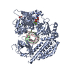
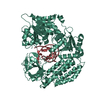
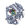
| Entry | Database: PDB / ID: 6iqr | ||||||
|---|---|---|---|---|---|---|---|
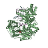
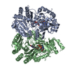
| Title | Crystal structure of Prc with S452I and L252Y mutations | ||||||
 Components Components | Tail-specific protease | ||||||
 Keywords Keywords | HYDROLASE / protein quality control / peptidoglycan remodeling | ||||||
| Function / homology |  Function and homology information Function and homology informationsymbiont-mediated evasion of recognition by host complement / C-terminal processing peptidase / protein catabolic process / outer membrane-bounded periplasmic space / endopeptidase activity / serine-type endopeptidase activity / response to antibiotic / signal transduction / proteolysis / plasma membrane Similarity search - Function | ||||||
| Biological species |  | ||||||
| Method |  X-RAY DIFFRACTION / SYNCHROTRON / MOLECULAR REPLACEMENT / Resolution: 3.42 Å X-RAY DIFFRACTION / SYNCHROTRON / MOLECULAR REPLACEMENT / Resolution: 3.42 Å | ||||||
 Authors Authors | Chueh, C.K. / Chang, C.I. | ||||||
| Funding support |  Taiwan, 1items Taiwan, 1items
| ||||||
 Citation Citation | Journal: Mbio / Year: 2019 Title: Structural Basis for the Differential Regulatory Roles of the PDZ Domain in C-Terminal Processing Proteases. Authors: Chueh, C.K. / Som, N. / Ke, L.C. / Ho, M.R. / Reddy, M. / Chang, C.I. | ||||||
| History |
|
- Structure visualization
| Structure viewer | Molecule: MolmilJmol/JSmol |
|---|
- Downloads & links
-Download
| PDBx/mmCIF format | 6iqr.cif.gz | 260 KB | Display | PDBx/mmCIF format |
|---|---|---|---|---|
| PDB format | pdb6iqr.ent.gz | 209.9 KB | Display | PDB format |
| PDBx/mmJSON format | 6iqr.json.gz | Tree view | PDBx/mmJSON format | |
| Others |  Other downloads Other downloads |
-Validation report
| Arichive directory | https://data.pdbj.org/pub/pdb/validation_reports/iq/6iqrftp://data.pdbj.org/pub/pdb/validation_reports/iq/6iqr | HTTPS FTP |
|---|
-Related structure data
| Related structure data |  6iqqC  6iqsC  6iquC  5wqlS S: Starting model for refinement C: citing same article ( |
|---|---|
| Similar structure data |


-Links
 PDBj
PDBj- Assembly
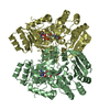
| Deposited unit | 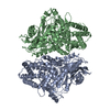
| ||||||||||||||||||
|---|---|---|---|---|---|---|---|---|---|---|---|---|---|---|---|---|---|---|---|
| 1 | 
| ||||||||||||||||||
| 2 | 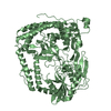
| ||||||||||||||||||
| Unit cell |
| ||||||||||||||||||
| Noncrystallographic symmetry (NCS) | NCS domain:
NCS domain segments: Component-ID: _ / Ens-ID: 1 / Beg auth comp-ID: ILE / Beg label comp-ID: ILE / End auth comp-ID: GLU / End label comp-ID: GLU / Refine code: _ / Auth seq-ID: 32 - 670 / Label seq-ID: 32 - 670
|
-Components
| #1: Protein | Mass: 77670.945 Da / Num. of mol.: 2 / Mutation: L252Y/S452I Source method: isolated from a genetically manipulated source Source: (gene. exp.) Strain: K12 / Gene: prc, tsp, b1830, JW1819 / Production host: References: UniProt: P23865, C-terminal processing peptidase #2: Water | ChemComp-HOH / |  Mass: 18.015 Da / Num. of mol.: 2 / Source method: isolated from a natural source / Formula: H2O Mass: 18.015 Da / Num. of mol.: 2 / Source method: isolated from a natural source / Formula: H2O |
|---|
-Experimental details
-Experiment
| Experiment | Method: X-RAY DIFFRACTION / Number of used crystals: 1 |
|---|
- Sample preparation
| Crystal | Density Matthews: 3.68 Å3/Da / Density % sol: 66.8 % |
|---|---|
| Crystal grow | Temperature: 295 K / Method: vapor diffusion, sitting drop / Details: PEG3350, sodium thyocinate |
-Data collection
| Diffraction | Mean temperature: 110 K / Serial crystal experiment: N |
|---|---|
| Diffraction source | Source: SYNCHROTRON / Site: NSRRC / Beamline: BL15A1 / Wavelength: 1 Å |
| Detector | Type: RAYONIX MX300HE / Detector: CCD / Date: Jun 7, 2018 Details: Vertically Collimating Premirror, LN2-Cooled Fixed-Exit Double Crystal Si(111) Monochromator , Toroidal Focusing Mirror |
| Radiation | Monochromator: LN2-Cooled, Fixed-Exit Double Crystal Monochromator Protocol: SINGLE WAVELENGTH / Monochromatic (M) / Laue (L): M / Scattering type: x-ray |
| Radiation wavelength | Wavelength: 1 Å / Relative weight: 1 |
| Reflection | Resolution: 3.42→50 Å / Num. obs: 31494 / % possible obs: 99.4 % / Redundancy: 4.3 % / Biso Wilson estimate: 44.65 Å2 / Rmerge(I) obs: 0.075 / Rpim(I) all: 0.069 / Rrim(I) all: 0.145 / Χ2: 0.968 / Net I/σ(I): 13.8 |
| Reflection shell | Resolution: 3.42→3.54 Å / Redundancy: 4.5 % / Rmerge(I) obs: 0.651 / Mean I/σ(I) obs: 2.1 / Num. unique obs: 3117 / CC1/2: 0.772 / Rpim(I) all: 0.446 / Rrim(I) all: 0.942 / Χ2: 0.95 / % possible all: 100 |
- Processing
| Software |
| ||||||||||||||||||||||||||||||||||||||||||||||||||||||||||||||||||||||||||||||||||||||||||||||||||||||||||||||||||||||||||||||||||||||||||||||||||||||||||||||||||||||||||||||||||||||
|---|---|---|---|---|---|---|---|---|---|---|---|---|---|---|---|---|---|---|---|---|---|---|---|---|---|---|---|---|---|---|---|---|---|---|---|---|---|---|---|---|---|---|---|---|---|---|---|---|---|---|---|---|---|---|---|---|---|---|---|---|---|---|---|---|---|---|---|---|---|---|---|---|---|---|---|---|---|---|---|---|---|---|---|---|---|---|---|---|---|---|---|---|---|---|---|---|---|---|---|---|---|---|---|---|---|---|---|---|---|---|---|---|---|---|---|---|---|---|---|---|---|---|---|---|---|---|---|---|---|---|---|---|---|---|---|---|---|---|---|---|---|---|---|---|---|---|---|---|---|---|---|---|---|---|---|---|---|---|---|---|---|---|---|---|---|---|---|---|---|---|---|---|---|---|---|---|---|---|---|---|---|---|---|
| Refinement | Method to determine structure: MOLECULAR REPLACEMENT Starting model: 5WQL Resolution: 3.42→29.97 Å / Cor.coef. Fo:Fc: 0.846 / Cor.coef. Fo:Fc free: 0.79 / SU B: 18.879 / SU ML: 0.336 / Cross valid method: THROUGHOUT / ESU R Free: 0.631 / Details: HYDROGENS HAVE BEEN ADDED IN THE RIDING POSITIONS
| ||||||||||||||||||||||||||||||||||||||||||||||||||||||||||||||||||||||||||||||||||||||||||||||||||||||||||||||||||||||||||||||||||||||||||||||||||||||||||||||||||||||||||||||||||||||
| Solvent computation | Ion probe radii: 0.8 Å / Shrinkage radii: 0.8 Å / VDW probe radii: 1.2 Å | ||||||||||||||||||||||||||||||||||||||||||||||||||||||||||||||||||||||||||||||||||||||||||||||||||||||||||||||||||||||||||||||||||||||||||||||||||||||||||||||||||||||||||||||||||||||
| Displacement parameters | Biso mean: 71.348 Å2
| ||||||||||||||||||||||||||||||||||||||||||||||||||||||||||||||||||||||||||||||||||||||||||||||||||||||||||||||||||||||||||||||||||||||||||||||||||||||||||||||||||||||||||||||||||||||
| Refinement step | Cycle: 1 / Resolution: 3.42→29.97 Å
| ||||||||||||||||||||||||||||||||||||||||||||||||||||||||||||||||||||||||||||||||||||||||||||||||||||||||||||||||||||||||||||||||||||||||||||||||||||||||||||||||||||||||||||||||||||||
| Refine LS restraints |
|