Mass: 18.015 Da / Num. of mol.: 493 / Source method: isolated from a natural source / Formula: H2O
Sequence details
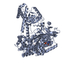
PROTEIN EXPRESSED WITH A C-TERMINAL HIS-TAG. PRIOR TO CRYSTALLISATION, UBII WAS SUBJECTED TO ...PROTEIN EXPRESSED WITH A C-TERMINAL HIS-TAG. PRIOR TO CRYSTALLISATION, UBII WAS SUBJECTED TO LIMITED PROTEOLYSIS WITH TRYPSIN. TWO CLEAVAGE SITES WERE IDENTIFIED BY MASS SPECTROMETRY TO BE LOCATED AT THE C-TERMINAL OF RESIDUE LYS 364 and LYS 365. THE C-TERMINAL FRAGMENT (34 RESIDUES) CONTAINING THE HIS-TAG WAS REMOVED BY NI-NTA PURIFICATION.
-
Experimental details
-
Experiment
Experiment
Method: X-RAY DIFFRACTION / Number of used crystals: 2
Protocol: SINGLE WAVELENGTH / Monochromatic (M) / Laue (L): M / Scattering type: x-ray
Radiation wavelength
ID
Wavelength (Å)
Relative weight
1
0.98011
1
2
0.97918
1
Reflection
Resolution: 2→46.98 Å / Num. obs: 58014 / % possible obs: 99.5 % / Observed criterion σ(I): 3 / Redundancy: 5.2 % / Biso Wilson estimate: 35.15 Å2 / Rmerge(I) obs: 0.0666 / Net I/σ(I): 14.5
-
Processing
Software
Name
Version
Classification
PHASER
2.5.2
phasing
PHENIX
(phenix.refine: 1.8.2_1309)
refinement
XDS
datareduction
XSCALE
datascaling
Refinement
Method to determine structure: SAD then molecular replacement Resolution: 2→46.98 Å / SU ML: 0.2 / σ(F): 2 / Phase error: 19.78 / Stereochemistry target values: ML Details: Model calculated from experimental phases derived by SAD with selenomethionylated protein. Data used for SAD phasing stored in crystal2 dataset. Data for MR and refinement to 2.0 A ...Details: Model calculated from experimental phases derived by SAD with selenomethionylated protein. Data used for SAD phasing stored in crystal2 dataset. Data for MR and refinement to 2.0 A resolution stored in crystal1 dataset'
Rfactor
Num. reflection
% reflection
Selection details
Rfree
0.1981
2894
5 %
RANDOM
Rwork
0.1625
-
-
-
obs
0.1643
57894
99.85 %
-
all
-
58014
-
-
Solvent computation
Shrinkage radii: 1.2 Å / VDW probe radii: 1.3 Å / Solvent model: FLAT BULK SOLVENT MODEL
Refine analyze
Luzzati coordinate error obs: 0.22 Å
Refinement step
Cycle: LAST / Resolution: 2→46.98 Å
Protein
Nucleic acid
Ligand
Solvent
Total
Num. atoms
5495
0
53
493
6041
Refine LS restraints
Refine-ID
Type
Dev ideal
Number
X-RAY DIFFRACTION
f_bond_d
0.012
5787
X-RAY DIFFRACTION
f_angle_d
1.253
7840
X-RAY DIFFRACTION
f_dihedral_angle_d
14.102
2109
X-RAY DIFFRACTION
f_chiral_restr
0.055
848
X-RAY DIFFRACTION
f_plane_restr
0.008
1028
LS refinement shell
Refine-ID: X-RAY DIFFRACTION / Total num. of bins used: 21
Resolution (Å)
Rfactor Rfree
Num. reflection Rfree
Rfactor Rwork
Num. reflection Rwork
% reflection obs (%)
2-2.0328
0.2997
133
0.2493
2540
99
2.0328-2.0678
0.2705
136
0.2275
2589
100
2.0678-2.1054
0.2522
136
0.211
2583
100
2.1054-2.1459
0.2677
137
0.2014
2595
100
2.1459-2.1897
0.2437
135
0.1888
2567
100
2.1897-2.2374
0.2111
138
0.1865
2623
100
2.2374-2.2894
0.2382
136
0.1848
2590
100
2.2894-2.3467
0.2528
136
0.1738
2570
100
2.3467-2.4101
0.1992
137
0.1748
2607
100
2.4101-2.481
0.1991
138
0.1674
2622
100
2.481-2.5611
0.2367
137
0.1736
2599
100
2.5611-2.6526
0.2249
137
0.1738
2607
100
2.6526-2.7588
0.2177
137
0.1714
2600
100
2.7588-2.8844
0.2099
138
0.1706
2616
100
2.8844-3.0364
0.2064
137
0.1665
2610
100
3.0364-3.2266
0.1788
138
0.1552
2625
100
3.2266-3.4756
0.1787
139
0.1498
2637
100
3.4756-3.8253
0.1863
139
0.1464
2651
100
3.8253-4.3785
0.1459
140
0.1417
2653
100
4.3785-5.515
0.1867
143
0.1411
2708
100
5.515-46.9944
0.1983
147
0.1679
2808
99
Refinement TLS params.
Method: refined / Refine-ID: X-RAY DIFFRACTION
ID
L11 (°2)
L12 (°2)
L13 (°2)
L22 (°2)
L23 (°2)
L33 (°2)
S11 (Å °)
S12 (Å °)
S13 (Å °)
S21 (Å °)
S22 (Å °)
S23 (Å °)
S31 (Å °)
S32 (Å °)
S33 (Å °)
T11 (Å2)
T12 (Å2)
T13 (Å2)
T22 (Å2)
T23 (Å2)
T33 (Å2)
Origin x (Å)
Origin y (Å)
Origin z (Å)
1
1.6336
0.4764
-0.131
3.6789
0.8424
1.6718
-0.0277
-0.2107
-0.0327
0.5928
-0.0479
0.3671
0.1739
-0.028
0.0773
0.222
0.0517
0.0275
0.2624
-0.0176
0.2322
-18.5495
17.7978
-24.0577
2
0.9513
0.5599
0.2473
3.3466
1.1176
1.1813
-0.0409
-0.1299
0.0181
0.2274
0.09
-0.0016
0.1189
0.0505
-0.037
0.1541
0.0498
0.0042
0.2002
0.021
0.1659
-9.555
16.019
-30.5627
3
2.7833
-0.9941
-0.5366
1.5306
0.3147
1.4978
0.1162
0.6736
0.1588
-0.4243
-0.1272
-0.1247
-0.2085
-0.0962
-0.0124
0.3786
-0.1137
-0.0185
0.3848
0.0591
0.2188
7.8083
24.744
-61.6267
4
2.6226
-0.811
0.3436
1.2455
0.0757
0.6926
0.0654
0.3448
-0.0337
-0.2374
-0.0227
0.0086
-0.0694
-0.0861
-0.0506
0.2853
-0.0517
0.005
0.2622
0.0196
0.1644
0.8931
18.636
-54.9824
Refinement TLS group
ID
Refine-ID
Refine TLS-ID
Selection details
1
X-RAY DIFFRACTION
1
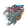
CHAINAAND (RESID1:140 )
2
X-RAY DIFFRACTION
2
CHAINAAND (RESID141:363 )
3
X-RAY DIFFRACTION
3
CHAINBAND (RESID2:140 )
4
X-RAY DIFFRACTION
4
CHAINBAND (RESID141:362 )
+
About Yorodumi
-
News
-
Feb 9, 2022. New format data for meta-information of EMDB entries
New format data for meta-information of EMDB entries
Version 3 of the EMDB header file is now the official format.
The previous official version 1.9 will be removed from the archive.
In the structure databanks used in Yorodumi, some data are registered as the other names, "COVID-19 virus" and "2019-nCoV". Here are the details of the virus and the list of structure data.
Jan 31, 2019. EMDB accession codes are about to change! (news from PDBe EMDB page)
EMDB accession codes are about to change! (news from PDBe EMDB page)
The allocation of 4 digits for EMDB accession codes will soon come to an end. Whilst these codes will remain in use, new EMDB accession codes will include an additional digit and will expand incrementally as the available range of codes is exhausted. The current 4-digit format prefixed with “EMD-” (i.e. EMD-XXXX) will advance to a 5-digit format (i.e. EMD-XXXXX), and so on. It is currently estimated that the 4-digit codes will be depleted around Spring 2019, at which point the 5-digit format will come into force.
The EM Navigator/Yorodumi systems omit the EMD- prefix.
Related info.:Q: What is EMD? / ID/Accession-code notation in Yorodumi/EM Navigator
Yorodumi is a browser for structure data from EMDB, PDB, SASBDB, etc.
This page is also the successor to EM Navigator detail page, and also detail information page/front-end page for Omokage search.
The word "yorodu" (or yorozu) is an old Japanese word meaning "ten thousand". "mi" (miru) is to see.
Related info.:EMDB / PDB / SASBDB / Comparison of 3 databanks / Yorodumi Search / Aug 31, 2016. New EM Navigator & Yorodumi / Yorodumi Papers / Jmol/JSmol / Function and homology information / Changes in new EM Navigator and Yorodumi
 Movie
Movie Controller

 Yorodumi
 Open data
 Basic information
 Components
 Keywords
 Function and homology information
 X-RAY DIFFRACTION /
  Authors
 Citation
 Structure visualization
 Downloads & links
 Other downloads


 PDBj
PDBj Assembly


 Mass: 92.094 Da / Num. of mol.: 7 / Source method: obtained synthetically / Formula: C3H8O3
 Mass: 35.453 Da / Num. of mol.: 4 / Source method: obtained synthetically / Formula: Cl
 Mass: 106.120 Da / Num. of mol.: 1 / Source method: obtained synthetically / Formula: C4H10O3
 Mass: 18.015 Da / Num. of mol.: 493 / Source method: isolated from a natural source / Formula: H2O
 Sample preparation
 Processing