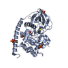
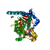
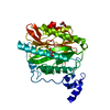
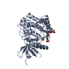
Entry Database : PDB / ID : 6s1sTitle Crystal structure of AmpC from Pseudomonas aeruginosa in complex with [3-(2-carboxyvinyl)phenyl]boronic acid] inhibitor Beta-lactamase Keywords / / / Function / homology Function Domain/homology Component
/ / / / / / / / / / / / / / / / / / / / / / / / Biological species Pseudomonas aeruginosa (bacteria)Method / / / Resolution : 1.78 Å Authors Kekez, I. / Vicario, M. / Bellio, P. / Tosoni, E. / Celenza, G. / Blazquez, J. / Tondi, D. / Cendron, L. Journal : Antibiotics / Year : 2019Title : Phenylboronic Acids Probing Molecular Recognition against Class A and Class C beta-lactamases.Authors : Linciano, P. / Vicario, M. / Kekez, I. / Bellio, P. / Celenza, G. / Martin-Blecua, I. / Blazquez, J. / Cendron, L. / Tondi, D. History Deposition Jun 19, 2019 Deposition site / Processing site Revision 1.0 Oct 9, 2019 Provider / Type Revision 1.1 Jan 24, 2024 Group / Database references / Refinement descriptionCategory chem_comp_atom / chem_comp_bond ... chem_comp_atom / chem_comp_bond / database_2 / pdbx_initial_refinement_model Item / _database_2.pdbx_database_accessionRevision 1.2 Oct 9, 2024 Group / Category / pdbx_modification_feature / Item
Show all Show less
 Movie
Movie Controller

 Yorodumi
 Open data
 Basic information
 Components
 Keywords
 Function and homology information
 Pseudomonas aeruginosa (bacteria)
 X-RAY DIFFRACTION /
  Authors
 Citation
 Structure visualization
 Downloads & links
 Other downloads
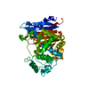


 PDBj
PDBj
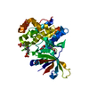
 Assembly
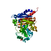
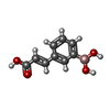
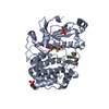
 Mass: 191.976 Da / Num. of mol.: 1 / Source method: obtained synthetically / Formula: C9H9BO4 / Feature type: SUBJECT OF INVESTIGATION
 Mass: 94.971 Da / Num. of mol.: 1 / Source method: obtained synthetically / Formula: PO4
 Mass: 18.015 Da / Num. of mol.: 131 / Source method: isolated from a natural source / Formula: H2O
 Sample preparation
 / Beamline: ID23-1 / Wavelength: 0.97779 Å
 Processing