 Movie
Movie Controller

[English] 日本語
 Yorodumi
- PDB-3af0: Pantothenate kinase from Mycobacterium tuberculosis (MtPanK) in c... -
+ Open data

- Basic information
| Entry | Database: PDB / ID: 3af0 | ||||||
|---|---|---|---|---|---|---|---|
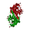
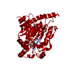
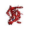
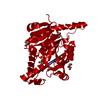
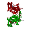
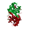
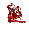
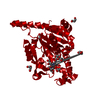
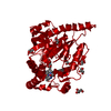
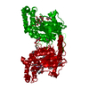
| Title | Pantothenate kinase from Mycobacterium tuberculosis (MtPanK) in complex with GDP and Pantothenate | ||||||
 Components Components | Pantothenate kinase | ||||||
 Keywords Keywords | TRANSFERASE / Homodimer / COA biosynthesis / Nucleotide binding / ATP-binding / Kinase / Nucleotide-binding | ||||||
| Function / homology |  Function and homology information Function and homology informationpantothenate kinase / pantothenate kinase activity / coenzyme A biosynthetic process / ATP binding / plasma membrane / cytoplasm Similarity search - Function | ||||||
| Biological species |   Mycobacterium tuberculosis (bacteria) Mycobacterium tuberculosis (bacteria) | ||||||
| Method |  X-RAY DIFFRACTION / MOLECULAR REPLACEMENT / Resolution: 2.5 Å X-RAY DIFFRACTION / MOLECULAR REPLACEMENT / Resolution: 2.5 Å | ||||||
 Authors Authors | Chetnani, B. / Kumar, P. / Surolia, A. / Vijayan, M. | ||||||
 Citation Citation | Journal: J.Mol.Biol. / Year: 2010 Title: M. tuberculosis pantothenate kinase: dual substrate specificity and unusual changes in ligand locations Authors: Chetnani, B. / Kumar, P. / Surolia, A. / Vijayan, M. | ||||||
| History |
|
- Structure visualization
| Structure viewer | Molecule: MolmilJmol/JSmol |
|---|
- Downloads & links
-Download
| PDBx/mmCIF format | 3af0.cif.gz | 83.8 KB | Display | PDBx/mmCIF format |
|---|---|---|---|---|
| PDB format | pdb3af0.ent.gz | 62 KB | Display | PDB format |
| PDBx/mmJSON format | 3af0.json.gz | Tree view | PDBx/mmJSON format | |
| Others |  Other downloads Other downloads |
-Validation report
| Arichive directory | https://data.pdbj.org/pub/pdb/validation_reports/af/3af0ftp://data.pdbj.org/pub/pdb/validation_reports/af/3af0 | HTTPS FTP |
|---|
-Related structure data
| Related structure data | 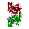 3aezC 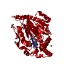 3af1C  3af2C 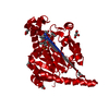 3af3C 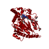 3af4C  2gevS C: citing same article ( S: Starting model for refinement |
|---|---|
| Similar structure data |


-Links
 PDBj
PDBj- Assembly
| Deposited unit | 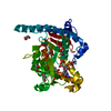
| ||||||||
|---|---|---|---|---|---|---|---|---|---|
| 1 | 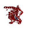
| ||||||||
| Unit cell |
|
-Components
-Protein , 1 types, 1 molecules A
| #1: Protein | Mass: 35704.848 Da / Num. of mol.: 1 Source method: isolated from a genetically manipulated source Source: (gene. exp.) Mycobacterium tuberculosis (bacteria) / Strain: H37Rv / Gene: coaA, Rv1092c / Plasmid: PET-28a(+) (NOVAGEN) / Production host: References: UniProt: P63810, UniProt: P9WPA7*PLUS, pantothenate kinase |
|---|
-Non-polymers , 5 types, 187 molecules 


| #2: Chemical | ChemComp-GDP /  Type: RNA linking / Mass: 443.201 Da / Num. of mol.: 1 / Source method: obtained synthetically / Formula: C10H15N5O11P2 / Comment: GDP, energy-carrying molecule*YM Type: RNA linking / Mass: 443.201 Da / Num. of mol.: 1 / Source method: obtained synthetically / Formula: C10H15N5O11P2 / Comment: GDP, energy-carrying molecule*YM | ||
|---|---|---|---|
| #3: Chemical | ChemComp-PAU /  Type: peptide-like / Mass: 219.235 Da / Num. of mol.: 1 / Source method: obtained synthetically / Formula: C9H17NO5 Type: peptide-like / Mass: 219.235 Da / Num. of mol.: 1 / Source method: obtained synthetically / Formula: C9H17NO5 | ||
| #4: Chemical | ChemComp-CL /  Mass: 35.453 Da / Num. of mol.: 1 / Source method: obtained synthetically / Formula: Cl Mass: 35.453 Da / Num. of mol.: 1 / Source method: obtained synthetically / Formula: Cl | ||
| #5: Chemical | ChemComp-GOL /  Mass: 92.094 Da / Num. of mol.: 9 / Source method: obtained synthetically / Formula: C3H8O3 Mass: 92.094 Da / Num. of mol.: 9 / Source method: obtained synthetically / Formula: C3H8O3#6: Water | ChemComp-HOH / | Mass: 18.015 Da / Num. of mol.: 175 / Source method: isolated from a natural source / Formula: H2O |
-Experimental details
-Experiment
| Experiment | Method: X-RAY DIFFRACTION / Number of used crystals: 1 |
|---|
- Sample preparation
| Crystal | Density Matthews: 4.01 Å3/Da / Density % sol: 69.31 % |
|---|---|
| Crystal grow | Temperature: 293 K / Method: vapor diffusion, hanging drop / pH: 6.5 Details: 1.4-1.8M trisodium citrate, 0.05-0.1M sodium acetate, 10% glycerol, pH 6.5, VAPOR DIFFUSION, HANGING DROP, temperature 293K |
-Data collection
| Diffraction | Mean temperature: 100 K |
|---|---|
| Diffraction source | Source: ROTATING ANODE / Type: BRUKER AXS MICROSTAR / Wavelength: 1.54 Å |
| Detector | Type: MAR scanner 345 mm plate / Detector: IMAGE PLATE / Date: May 19, 2009 / Details: Mirrors |
| Radiation | Monochromator: OSMIC MIRROR / Protocol: SINGLE WAVELENGTH / Monochromatic (M) / Laue (L): M / Scattering type: x-ray |
| Radiation wavelength | Wavelength: 1.54 Å / Relative weight: 1 |
| Reflection | Resolution: 2.5→40 Å / Num. obs: 20208 / % possible obs: 99.9 % / Observed criterion σ(I): 0 / Redundancy: 3.9 % / Biso Wilson estimate: 65.7 Å2 / Rmerge(I) obs: 0.063 / Net I/σ(I): 13 |
| Reflection shell | Resolution: 2.5→2.64 Å / Redundancy: 3.6 % / Rmerge(I) obs: 0.548 / Mean I/σ(I) obs: 2 / Num. unique all: 2893 / % possible all: 99.8 |
- Processing
| Software |
| |||||||||||||||||||||||||||||||||||||||||||||||||||||||||||||||||
|---|---|---|---|---|---|---|---|---|---|---|---|---|---|---|---|---|---|---|---|---|---|---|---|---|---|---|---|---|---|---|---|---|---|---|---|---|---|---|---|---|---|---|---|---|---|---|---|---|---|---|---|---|---|---|---|---|---|---|---|---|---|---|---|---|---|---|
| Refinement | Method to determine structure: MOLECULAR REPLACEMENT Starting model: 2GEV Resolution: 2.5→34.28 Å / Cor.coef. Fo:Fc: 0.949 / Cor.coef. Fo:Fc free: 0.926 / Cross valid method: THROUGHOUT / σ(F): 0 / ESU R: 0.304 / ESU R Free: 0.244 / Stereochemistry target values: MAXIMUM LIKELIHOOD / Details: BULK SOLVENT MODEL USED
| |||||||||||||||||||||||||||||||||||||||||||||||||||||||||||||||||
| Solvent computation | Ion probe radii: 0.8 Å / Shrinkage radii: 0.8 Å / VDW probe radii: 1.4 Å / Solvent model: MASK | |||||||||||||||||||||||||||||||||||||||||||||||||||||||||||||||||
| Displacement parameters | Biso mean: 49.796 Å2
| |||||||||||||||||||||||||||||||||||||||||||||||||||||||||||||||||
| Refinement step | Cycle: LAST / Resolution: 2.5→34.28 Å
| |||||||||||||||||||||||||||||||||||||||||||||||||||||||||||||||||
| Refine LS restraints |
| |||||||||||||||||||||||||||||||||||||||||||||||||||||||||||||||||
| LS refinement shell | Resolution: 2.5→2.565 Å / Total num. of bins used: 20
|
