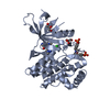
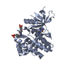
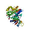
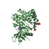
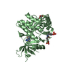
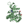
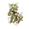
Entry Database : PDB / ID : 6n78Title Structure of the human JAK1 kinase domain with compound 21 Tyrosine-protein kinase JAK1 Keywords / / / / / / Function / homology Function Domain/homology Component
/ / / / / / / / / / / / / / / / / / / / / / / / / / / / / / / / / / / / / / / / / / / / / / / / / / / / / / / / / / / / / / / / / / / / / / / / / / / / / / / / / / / / / / / / / / / / / / / / / / / / / / / / / / / / / / / / / / / / / / / / / / / / / / Biological species Homo sapiens (human)Method / / Resolution : 1.83 Å Authors Lupardus, P.J. / Brown, D. Journal : Bioorg.Med.Chem.Lett. / Year : 2019Title : Discovery of a class of highly potent Janus Kinase 1/2 (JAK1/2) inhibitors demonstrating effective cell-based blockade of IL-13 signaling.Authors: Zak, M. / Hanan, E.J. / Lupardus, P. / Brown, D.G. / Robinson, C. / Siu, M. / Lyssikatos, J.P. / Romero, F.A. / Zhao, G. / Kellar, T. / Mendonca, R. / Ray, N.C. / Goodacre, S.C. / Crackett, ... Authors : Zak, M. / Hanan, E.J. / Lupardus, P. / Brown, D.G. / Robinson, C. / Siu, M. / Lyssikatos, J.P. / Romero, F.A. / Zhao, G. / Kellar, T. / Mendonca, R. / Ray, N.C. / Goodacre, S.C. / Crackett, P.H. / McLean, N. / Hurley, C.A. / Yuen, P.W. / Cheng, Y.X. / Liu, X. / Liimatta, M. / Kohli, P.B. / Nonomiya, J. / Salmon, G. / Buckley, G. / Lloyd, J. / Gibbons, P. / Ghilardi, N. / Kenny, J.R. / Johnson, A. History Deposition Nov 27, 2018 Deposition site / Processing site Revision 1.0 Apr 24, 2019 Provider / Type Revision 1.1 May 15, 2019 Group / Database references / Category Item _citation.journal_abbrev / _citation.journal_volume ... _citation.journal_abbrev / _citation.journal_volume / _citation.page_first / _citation.page_last Revision 1.2 Nov 6, 2024 Group / Database references / Structure summaryCategory chem_comp_atom / chem_comp_bond ... chem_comp_atom / chem_comp_bond / database_2 / pdbx_entry_details / pdbx_modification_feature Item / _database_2.pdbx_database_accession
Show all Show less
 Movie
Movie Controller


 Open data
 Basic information
 Components
 Keywords
 Function and homology information
 Homo sapiens (human)
 X-RAY DIFFRACTION /
  Authors
 Citation
 Structure visualization
 Downloads & links
 Other downloads
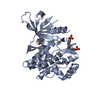


 PDBj
PDBj


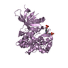
 Assembly

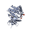
 Spodoptera frugiperda (fall armyworm)
 Mass: 418.785 Da / Num. of mol.: 1 / Source method: obtained synthetically / Formula: C18H13ClF2N6O2
 Mass: 92.094 Da / Num. of mol.: 1 / Source method: obtained synthetically / Formula: C3H8O3
 Mass: 18.015 Da / Num. of mol.: 146 / Source method: isolated from a natural source / Formula: H2O
 Sample preparation
 / Beamline: I04-1 / Wavelength: 0.9282 Å
 Processing