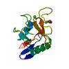
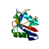
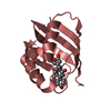
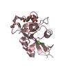
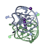
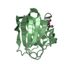
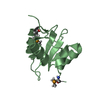
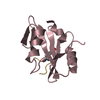
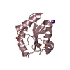
Evidence: From known literature, active molecule is composed of four trimeric units - trimeric unit is composed of N-terminal domain, collagen-like helix, alpha-helical coiled-coil and 3 carbohydrate ...Evidence: From known literature, active molecule is composed of four trimeric units - trimeric unit is composed of N-terminal domain, collagen-like helix, alpha-helical coiled-coil and 3 carbohydrate recognition domains. As the structure only contains a carbohydrate recognition domain, it is only a monomer.
Type
Name
Symmetry operation
Number
identity operation
1_555
x,y,z
1
Buried area
500 Å2
ΔGint
-23 kcal/mol
Surface area
6330 Å2
Method
PISA
Unit cell
Length a, b, c (Å)
50.278, 50.278, 52.192
Angle α, β, γ (deg.)
90.000, 90.000, 90.000
Int Tables number
78
Space group name H-M
P43
-
Components
#1: Protein
Conglutinin
Mass: 14082.562 Da / Num. of mol.: 1 / Fragment: carbohydrate recognition domain Source method: isolated from a genetically manipulated source Source: (gene. exp.) Bos taurus (domestic cattle) / Gene: CGN1 / Production host: Escherichia coli BL21(DE3) (bacteria) / References: UniProt: P23805
Resolution: 1→50 Å / Cor.coef. Fo:Fc: 0.975 / Cor.coef. Fo:Fc free: 0.968 / SU B: 0.413 / SU ML: 0.01 / SU R Cruickshank DPI: 0.0177 / Cross valid method: THROUGHOUT / σ(F): 0 / ESU R: 0.018 / ESU R Free: 0.018 Details: HYDROGENS HAVE BEEN ADDED IN THE RIDING POSITIONS U VALUES : REFINED INDIVIDUALLY
Rfactor
Num. reflection
% reflection
Selection details
Rfree
0.14
3523
5 %
RANDOM
Rwork
0.1239
-
-
-
obs
0.1247
66521
99.46 %
-
Solvent computation
Ion probe radii: 0.8 Å / Shrinkage radii: 0.8 Å / VDW probe radii: 1.2 Å
In the structure databanks used in Yorodumi, some data are registered as the other names, "COVID-19 virus" and "2019-nCoV". Here are the details of the virus and the list of structure data.
Jan 31, 2019. EMDB accession codes are about to change! (news from PDBe EMDB page)
EMDB accession codes are about to change! (news from PDBe EMDB page)
The allocation of 4 digits for EMDB accession codes will soon come to an end. Whilst these codes will remain in use, new EMDB accession codes will include an additional digit and will expand incrementally as the available range of codes is exhausted. The current 4-digit format prefixed with “EMD-” (i.e. EMD-XXXX) will advance to a 5-digit format (i.e. EMD-XXXXX), and so on. It is currently estimated that the 4-digit codes will be depleted around Spring 2019, at which point the 5-digit format will come into force.
The EM Navigator/Yorodumi systems omit the EMD- prefix.
Related info.:Q: What is EMD? / ID/Accession-code notation in Yorodumi/EM Navigator
Yorodumi is a browser for structure data from EMDB, PDB, SASBDB, etc.
This page is also the successor to EM Navigator detail page, and also detail information page/front-end page for Omokage search.
The word "yorodu" (or yorozu) is an old Japanese word meaning "ten thousand". "mi" (miru) is to see.
Related info.:EMDB / PDB / SASBDB / Comparison of 3 databanks / Yorodumi Search / Aug 31, 2016. New EM Navigator & Yorodumi / Yorodumi Papers / Jmol/JSmol / Function and homology information / Changes in new EM Navigator and Yorodumi
 Movie
Movie Controller

 Yorodumi
 Open data
 Basic information
 Components
 Keywords
 Function and homology information
 X-RAY DIFFRACTION /
  Authors
 Citation
 Structure visualization
 Downloads & links
 Other downloads


 PDBj
PDBj


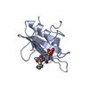
 Assembly


 Mass: 40.078 Da / Num. of mol.: 2 / Source method: obtained synthetically / Formula: Ca / Feature type: SUBJECT OF INVESTIGATION
 Type: D-saccharide / Mass: 301.188 Da / Num. of mol.: 1
 Mass: 18.015 Da / Num. of mol.: 173 / Source method: isolated from a natural source / Formula: H2O
 Sample preparation
 / Beamline: I04 / Wavelength: 0.827 Å
 Processing