 Movie
Movie Controller


+ Open data

- Basic information
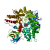
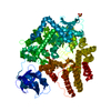
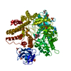
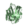
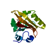
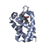
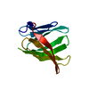
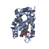
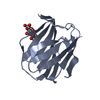
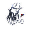
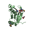
| Entry | Database: PDB / ID: 4a42 | ||||||
|---|---|---|---|---|---|---|---|
| Title | CpGH89CBM32-6 produced by Clostridium perfringens | ||||||
 Components Components | ALPHA-N-ACETYLGLUCOSAMINIDASE FAMILY PROTEIN | ||||||
 Keywords Keywords | HYDROLASE / FAMILY 89 GLYCOSIDE HYDROLASE / FAMILY 32 CARBOHYDRATE-BINDING MODULE / CPF_0859 | ||||||
| Function / homology |  Function and homology information Function and homology informationhydrolase activity, acting on glycosyl bonds / hydrolase activity, hydrolyzing O-glycosyl compounds / carbohydrate metabolic process / metal ion binding Similarity search - Function | ||||||
| Biological species |   CLOSTRIDIUM PERFRINGENS (bacteria) CLOSTRIDIUM PERFRINGENS (bacteria) | ||||||
| Method |  X-RAY DIFFRACTION / SYNCHROTRON / SAD / Resolution: 1.55 Å X-RAY DIFFRACTION / SYNCHROTRON / SAD / Resolution: 1.55 Å | ||||||
 Authors Authors | Ficko-Blean, E. / Stuart, C.P. / Suits, M.D. / Cid, M. / Tessier, M. / Woods, R.J. / Boraston, A.B. | ||||||
 Citation Citation | Journal: Plos One / Year: 2012 Title: Carbohydrate Recognition by an Architecturally Complex Alpha-N-Acetylglucosaminidase from Clostridium Perfringens. Authors: Ficko-Blean, E. / Stuart, C.P. / Suits, M.D. / Cid, M. / Tessier, M. / Woods, R.J. / Boraston, A.B. | ||||||
| History |
|
- Structure visualization
| Structure viewer | Molecule: MolmilJmol/JSmol |
|---|
- Downloads & links
-Download
| PDBx/mmCIF format | 4a42.cif.gz | 122.1 KB | Display | PDBx/mmCIF format |
|---|---|---|---|---|
| PDB format | pdb4a42.ent.gz | 95.1 KB | Display | PDB format |
| PDBx/mmJSON format | 4a42.json.gz | Tree view | PDBx/mmJSON format | |
| Others |  Other downloads Other downloads |
-Validation report
| Arichive directory | https://data.pdbj.org/pub/pdb/validation_reports/a4/4a42ftp://data.pdbj.org/pub/pdb/validation_reports/a4/4a42 | HTTPS FTP |
|---|
-Related structure data
| Related structure data |  4a3zC  4a41C  4a44C  4a45C  4a6oC  4aaxC  4a43 C: citing same article ( |
|---|---|
| Similar structure data |


-Links
 PDBj
PDBj


- Assembly
| Deposited unit | 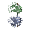
| ||||||||
|---|---|---|---|---|---|---|---|---|---|
| 1 | 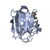
| ||||||||
| 2 | 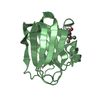
| ||||||||
| Unit cell |
|
-Components
| #1: Protein | Mass: 16823.033 Da / Num. of mol.: 2 / Fragment: CBM32-4, RESIDUES 1496-1621 Source method: isolated from a genetically manipulated source Source: (gene. exp.) CLOSTRIDIUM PERFRINGENS (bacteria) / Production host: References: UniProt: Q0TST1, UniProt: Q8XM24*PLUS, alpha-N-acetylglucosaminidase #2: Chemical |   Mass: 40.078 Da / Num. of mol.: 2 / Source method: obtained synthetically / Formula: Ca Mass: 40.078 Da / Num. of mol.: 2 / Source method: obtained synthetically / Formula: Ca#3: Water | ChemComp-HOH / |  Mass: 18.015 Da / Num. of mol.: 174 / Source method: isolated from a natural source / Formula: H2O Mass: 18.015 Da / Num. of mol.: 174 / Source method: isolated from a natural source / Formula: H2OHas protein modification | Y | Sequence details | SELENOMETH | |
|---|
-Experimental details
-Experiment
| Experiment | Method: X-RAY DIFFRACTION / Number of used crystals: 1 |
|---|
- Sample preparation
| Crystal | Density Matthews: 1.74 Å3/Da / Density % sol: 29.2 % / Description: NONE |
|---|
-Data collection
| Diffraction | Mean temperature: 100 K |
|---|---|
| Diffraction source | Source: SYNCHROTRON / Site: SSRL  / Beamline: BL9-2 / Wavelength: 0.979 / Beamline: BL9-2 / Wavelength: 0.979 |
| Detector | Type: MARMOSAIC 325 mm CCD / Detector: CCD |
| Radiation | Protocol: SINGLE WAVELENGTH / Monochromatic (M) / Laue (L): M / Scattering type: x-ray |
| Radiation wavelength | Wavelength: 0.979 Å / Relative weight: 1 |
| Reflection | Resolution: 1.55→30 Å / Num. obs: 33037 / % possible obs: 99.2 % / Observed criterion σ(I): 2 / Redundancy: 15.2 % / Biso Wilson estimate: 17.84 Å2 / Rmerge(I) obs: 0.05 / Net I/σ(I): 30.6 |
| Reflection shell | Resolution: 1.55→1.63 Å / Redundancy: 7.6 % / Rmerge(I) obs: 0.38 / Mean I/σ(I) obs: 7.6 / % possible all: 100 |
- Processing
| Software |
| |||||||||||||||||||||||||||||||||||||||||||||||||||||||||||||||||||||||||||||||||||||||||||
|---|---|---|---|---|---|---|---|---|---|---|---|---|---|---|---|---|---|---|---|---|---|---|---|---|---|---|---|---|---|---|---|---|---|---|---|---|---|---|---|---|---|---|---|---|---|---|---|---|---|---|---|---|---|---|---|---|---|---|---|---|---|---|---|---|---|---|---|---|---|---|---|---|---|---|---|---|---|---|---|---|---|---|---|---|---|---|---|---|---|---|---|---|
| Refinement | Method to determine structure: SAD Starting model: NONE Resolution: 1.55→28.234 Å / SU ML: 0.21 / σ(F): 0 / Phase error: 22.78 / Stereochemistry target values: ML Details: DISORDERED REGIONS WERE MODELED STEREOCHEMICALLY AND RESIDUES WITH POOR SIDE CHAIN DENSITY ARE PRESENTED AS STUBS.
| |||||||||||||||||||||||||||||||||||||||||||||||||||||||||||||||||||||||||||||||||||||||||||
| Solvent computation | Shrinkage radii: 0.9 Å / VDW probe radii: 1.11 Å / Solvent model: FLAT BULK SOLVENT MODEL / Bsol: 47.2 Å2 / ksol: 0.361 e/Å3 | |||||||||||||||||||||||||||||||||||||||||||||||||||||||||||||||||||||||||||||||||||||||||||
| Displacement parameters |
| |||||||||||||||||||||||||||||||||||||||||||||||||||||||||||||||||||||||||||||||||||||||||||
| Refinement step | Cycle: LAST / Resolution: 1.55→28.234 Å
| |||||||||||||||||||||||||||||||||||||||||||||||||||||||||||||||||||||||||||||||||||||||||||
| Refine LS restraints |
| |||||||||||||||||||||||||||||||||||||||||||||||||||||||||||||||||||||||||||||||||||||||||||
| LS refinement shell |
|
