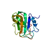
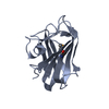
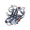
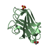
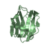
Hydrolases; Glycosylases; Glycosidases, i.e. enzymes that hydrolyse O- and S-glycosyl compounds / polysaccharide catabolic process / hydrolase activity, hydrolyzing O-glycosyl compounds Similarity search - Function
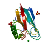
Glycoside hydrolase, family 8, conserved site / Glycosyl hydrolases family 8 signature. / NedA-like, galactose-binding domain / Glycoside hydrolase, family 8 / Glycosyl hydrolases family 8 / Coagulation factor 5/8 C-terminal domain, discoidin domain / Coagulation factors 5/8 type C domain (FA58C) profile. / F5/8 type C domain / Coagulation factor 5/8 C-terminal domain / Six-hairpin glycosidase-like superfamily ...Glycoside hydrolase, family 8, conserved site / Glycosyl hydrolases family 8 signature. / NedA-like, galactose-binding domain / Glycoside hydrolase, family 8 / Glycosyl hydrolases family 8 / Coagulation factor 5/8 C-terminal domain, discoidin domain / Coagulation factors 5/8 type C domain (FA58C) profile. / F5/8 type C domain / Coagulation factor 5/8 C-terminal domain / Six-hairpin glycosidase-like superfamily / Six-hairpin glycosidase superfamily / Galactose-binding domain-like / Fibronectin type III domain / Fibronectin type 3 domain / Fibronectin type-III domain profile. / Galactose-binding-like domain superfamily / Fibronectin type III / Fibronectin type III superfamily / Jelly Rolls / Immunoglobulin-like fold / Sandwich / Mainly Beta Similarity search - Domain/homology
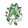
 Movie
Movie Controller

 Yorodumi
 Open data
 Basic information
 Components
 Keywords
 Function and homology information
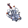
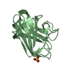
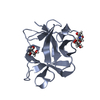
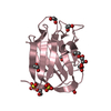
 Paenibacillus fukuinensis (bacteria)
 X-RAY DIFFRACTION /
  Authors
 Japan, 1items
  Citation
 Structure visualization
 Downloads & links
 Other downloads
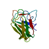


 PDBj
PDBj


 Assembly


 Mass: 62.068 Da / Num. of mol.: 18 / Source method: obtained synthetically / Formula: C2H6O2
 Mass: 96.063 Da / Num. of mol.: 4 / Source method: obtained synthetically / Formula: SO4
 Mass: 18.015 Da / Num. of mol.: 625 / Source method: isolated from a natural source / Formula: H2O
 Sample preparation
 Processing