 Movie
Movie Controller

[English] 日本語
 Yorodumi
- PDB-3ef4: Crystal structure of native pseudoazurin from Hyphomicrobium deni... -
+ Open data

- Basic information
| Entry | Database: PDB / ID: 3ef4 | ||||||
|---|---|---|---|---|---|---|---|
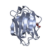
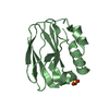
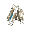
| Title | Crystal structure of native pseudoazurin from Hyphomicrobium denitrificans | ||||||
 Components Components | Blue copper protein | ||||||
 Keywords Keywords | ELECTRON TRANSPORT / copper / electron transfer / blue copper protein | ||||||
| Function / homology |  Function and homology information Function and homology information | ||||||
| Biological species |  Hyphomicrobium denitrificans (bacteria) Hyphomicrobium denitrificans (bacteria) | ||||||
| Method |  X-RAY DIFFRACTION / SYNCHROTRON / MOLECULAR REPLACEMENT / Resolution: 1.18 Å X-RAY DIFFRACTION / SYNCHROTRON / MOLECULAR REPLACEMENT / Resolution: 1.18 Å | ||||||
 Authors Authors | Hira, D. / Nojiri, M. / Suzuki, S. | ||||||
 Citation Citation | Journal: Acta Crystallogr.,Sect.D / Year: 2009 Title: Atomic resolution structure of pseudoazurin from the methylotrophic denitrifying bacterium Hyphomicrobium denitrificans: structural insights into its spectroscopic properties Authors: Hira, D. / Nojiri, M. / Suzuki, S. | ||||||
| History |
|
- Structure visualization
| Structure viewer | Molecule: MolmilJmol/JSmol |
|---|
- Downloads & links
-Download
| PDBx/mmCIF format | 3ef4.cif.gz | 181.6 KB | Display | PDBx/mmCIF format |
|---|---|---|---|---|
| PDB format | pdb3ef4.ent.gz | 144.3 KB | Display | PDB format |
| PDBx/mmJSON format | 3ef4.json.gz | Tree view | PDBx/mmJSON format | |
| Others |  Other downloads Other downloads |
-Validation report
| Arichive directory | https://data.pdbj.org/pub/pdb/validation_reports/ef/3ef4ftp://data.pdbj.org/pub/pdb/validation_reports/ef/3ef4 | HTTPS FTP |
|---|
-Related structure data
| Related structure data |  1adwS S: Starting model for refinement |
|---|---|
| Similar structure data |


-Links
 PDBj
PDBj- Assembly
| Deposited unit | 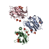
| |||||||||
|---|---|---|---|---|---|---|---|---|---|---|
| 1 | 
| |||||||||
| 2 | 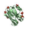
| |||||||||
| 3 | 
| |||||||||
| Unit cell |
| |||||||||
| Components on special symmetry positions |
|
-Components
| #1: Protein | Mass: 13590.666 Da / Num. of mol.: 3 / Source method: isolated from a natural source / Source: (natural) Hyphomicrobium denitrificans (bacteria) / References: UniProt: A7VL37#2: Chemical |   Mass: 63.546 Da / Num. of mol.: 3 / Source method: obtained synthetically / Formula: Cu Mass: 63.546 Da / Num. of mol.: 3 / Source method: obtained synthetically / Formula: Cu#3: Chemical | ChemComp-PO4 /   Mass: 94.971 Da / Num. of mol.: 15 / Source method: obtained synthetically / Formula: PO4 Mass: 94.971 Da / Num. of mol.: 15 / Source method: obtained synthetically / Formula: PO4#4: Water | ChemComp-HOH / |  Mass: 18.015 Da / Num. of mol.: 500 / Source method: isolated from a natural source / Formula: H2O Mass: 18.015 Da / Num. of mol.: 500 / Source method: isolated from a natural source / Formula: H2O |
|---|
-Experimental details
-Experiment
| Experiment | Method: X-RAY DIFFRACTION / Number of used crystals: 1 |
|---|
- Sample preparation
| Crystal | Density Matthews: 2.5 Å3/Da / Density % sol: 50.75 % |
|---|---|
| Crystal grow | Temperature: 277 K / Method: vapor diffusion, hanging drop / pH: 4 Details: 3.2M Ammonium Phosphate, 0.05M Potassium Phosphate, pH 4.0, VAPOR DIFFUSION, HANGING DROP, temperature 277K |
-Data collection
| Diffraction | Mean temperature: 100 K |
|---|---|
| Diffraction source | Source: SYNCHROTRON / Site: SPring-8  / Beamline: BL44XU / Wavelength: 0.9 Å / Beamline: BL44XU / Wavelength: 0.9 Å |
| Detector | Type: Bruker DIP-6040 / Detector: CCD / Date: Jun 13, 2006 |
| Radiation | Monochromator: double Si(111) / Protocol: SINGLE WAVELENGTH / Monochromatic (M) / Laue (L): M / Scattering type: x-ray |
| Radiation wavelength | Wavelength: 0.9 Å / Relative weight: 1 |
| Reflection | Resolution: 1.18→49.2 Å / Num. all: 132934 / Num. obs: 132934 / % possible obs: 95.8 % / Observed criterion σ(F): 0 / Observed criterion σ(I): 0 / Redundancy: 3.8 % / Rmerge(I) obs: 0.041 / Net I/σ(I): 43.59 |
| Reflection shell | Resolution: 1.18→1.22 Å / Redundancy: 2.9 % / Rmerge(I) obs: 0.49 / Mean I/σ(I) obs: 2.78 / % possible all: 85.2 |
- Processing
| Software |
| |||||||||||||||||||||||||
|---|---|---|---|---|---|---|---|---|---|---|---|---|---|---|---|---|---|---|---|---|---|---|---|---|---|---|
| Refinement | Method to determine structure: MOLECULAR REPLACEMENT Starting model: PDB ENTRY 1adw Resolution: 1.18→44.95 Å / Num. parameters: 32026 / Num. restraintsaints: 45026 / Cross valid method: FREE R / σ(F): 0 / Stereochemistry target values: Engh & Huber
| |||||||||||||||||||||||||
| Refine analyze | Num. disordered residues: 30 / Occupancy sum hydrogen: 2724 / Occupancy sum non hydrogen: 3402 | |||||||||||||||||||||||||
| Refinement step | Cycle: LAST / Resolution: 1.18→44.95 Å
| |||||||||||||||||||||||||
| Refine LS restraints |
|
