 Movie
Movie Controller

[English] 日本語
 Yorodumi
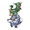
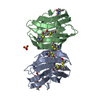
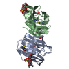
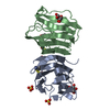
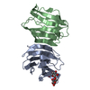
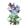
- PDB-4q26: Crystal Structure of Galectin-1 in Complex with N-Acetyllactosamine -
+ Open data

- Basic information
| Entry | Database: PDB / ID: 4q26 | |||||||||
|---|---|---|---|---|---|---|---|---|---|---|
| Title | Crystal Structure of Galectin-1 in Complex with N-Acetyllactosamine | |||||||||
 Components Components | Galectin-1 | |||||||||
 Keywords Keywords | SUGAR BINDING PROTEIN | |||||||||
| Function / homology |  Function and homology information Function and homology informationgalectin complex / lactose binding / negative regulation of T-helper 17 cell lineage commitment / myoblast differentiation / plasma cell differentiation / T cell costimulation / laminin binding / Post-translational protein phosphorylation / cell-cell adhesion / Regulation of Insulin-like Growth Factor (IGF) transport and uptake by Insulin-like Growth Factor Binding Proteins (IGFBPs) ...galectin complex / lactose binding / negative regulation of T-helper 17 cell lineage commitment / myoblast differentiation / plasma cell differentiation / T cell costimulation / laminin binding / Post-translational protein phosphorylation / cell-cell adhesion / Regulation of Insulin-like Growth Factor (IGF) transport and uptake by Insulin-like Growth Factor Binding Proteins (IGFBPs) / positive regulation of inflammatory response / extracellular matrix / regulation of apoptotic process / positive regulation of viral entry into host cell / positive regulation of canonical NF-kappaB signal transduction / positive regulation of apoptotic process / endoplasmic reticulum lumen / receptor ligand activity / apoptotic process / : / RNA binding / extracellular exosome / extracellular region / plasma membrane / cytoplasm / cytosol Similarity search - Function | |||||||||
| Biological species |  Homo sapiens (human) Homo sapiens (human) | |||||||||
| Method |  X-RAY DIFFRACTION / SYNCHROTRON / MOLECULAR REPLACEMENT / Resolution: 1.399 Å X-RAY DIFFRACTION / SYNCHROTRON / MOLECULAR REPLACEMENT / Resolution: 1.399 Å | |||||||||
 Authors Authors | Grimm, C. / Bertleff-Zieschang, N. | |||||||||
 Citation Citation | Journal: To be Published Title: Crystal Structure of Galectin-1 in Complex with N-Acetyllactosamine Authors: Grimm, C. / Bertleff-Zieschang, N. | |||||||||
| History |
|
- Structure visualization
| Structure viewer | Molecule: MolmilJmol/JSmol |
|---|
- Downloads & links
-Download
| PDBx/mmCIF format | 4q26.cif.gz | 330.4 KB | Display | PDBx/mmCIF format |
|---|---|---|---|---|
| PDB format | pdb4q26.ent.gz | 277.2 KB | Display | PDB format |
| PDBx/mmJSON format | 4q26.json.gz | Tree view | PDBx/mmJSON format | |
| Others |  Other downloads Other downloads |
-Validation report
| Arichive directory | https://data.pdbj.org/pub/pdb/validation_reports/q2/4q26ftp://data.pdbj.org/pub/pdb/validation_reports/q2/4q26 | HTTPS FTP |
|---|
-Related structure data
| Related structure data | |
|---|---|
| Similar structure data |


-Links
 PDBj
PDBj- Assembly
| Deposited unit | 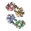
| ||||||||
|---|---|---|---|---|---|---|---|---|---|
| 1 | 
| ||||||||
| 2 | 
| ||||||||
| 3 | 
| ||||||||
| 4 | 
| ||||||||
| 5 | 
| ||||||||
| 6 | 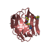
| ||||||||
| Unit cell |
| ||||||||
| Components on special symmetry positions |
|
-Components
| #1: Protein | Mass: 15016.079 Da / Num. of mol.: 4 Source method: isolated from a genetically manipulated source Source: (gene. exp.) Homo sapiens (human) / Gene: LGALS1 / Production host:  #2: Polysaccharide | beta-D-galactopyranose-(1-4)-2-acetamido-2-deoxy-alpha-D-glucopyranose / N-acetyl-alpha-lactosamine   Source method: isolated from a genetically manipulated source Details: oligosaccharide / References: N-acetyl-alpha-lactosamine #3: Chemical | ChemComp-GOL / |   Mass: 92.094 Da / Num. of mol.: 1 / Source method: obtained synthetically / Formula: C3H8O3 Mass: 92.094 Da / Num. of mol.: 1 / Source method: obtained synthetically / Formula: C3H8O3#4: Water | ChemComp-HOH / |  Mass: 18.015 Da / Num. of mol.: 495 / Source method: isolated from a natural source / Formula: H2O Mass: 18.015 Da / Num. of mol.: 495 / Source method: isolated from a natural source / Formula: H2OHas protein modification | Y | |
|---|
-Experimental details
-Experiment
| Experiment | Method: X-RAY DIFFRACTION / Number of used crystals: 1 |
|---|
- Sample preparation
| Crystal | Density Matthews: 2.74 Å3/Da / Density % sol: 55.15 % |
|---|---|
| Crystal grow | Temperature: 289 K / Method: vapor diffusion, hanging drop / pH: 7.5 Details: pH 7.5, VAPOR DIFFUSION, HANGING DROP, temperature 289K |
-Data collection
| Diffraction | Mean temperature: 100 K |
|---|---|
| Diffraction source | Source: SYNCHROTRON / Site: ESRF  / Beamline: ID23-1 / Beamline: ID23-1 |
| Detector | Type: DECTRIS PILATUS 6M-F / Detector: PIXEL |
| Radiation | Monochromator: Si(111) / Protocol: SINGLE WAVELENGTH / Monochromatic (M) / Laue (L): M / Scattering type: x-ray |
| Radiation wavelength | Relative weight: 1 |
| Reflection | Resolution: 1.399→48.023 Å / Num. all: 129682 / Num. obs: 129682 / % possible obs: 99.2 % / Observed criterion σ(I): 16.7 / Rsym value: 0.092 |
| Reflection shell | Highest resolution: 1.399 Å |
- Processing
| Software |
| |||||||||||||||||||||||||||||||||||||||||||||||||||||||||||||||||||||||||||||||||||||||||||||||||||||||||
|---|---|---|---|---|---|---|---|---|---|---|---|---|---|---|---|---|---|---|---|---|---|---|---|---|---|---|---|---|---|---|---|---|---|---|---|---|---|---|---|---|---|---|---|---|---|---|---|---|---|---|---|---|---|---|---|---|---|---|---|---|---|---|---|---|---|---|---|---|---|---|---|---|---|---|---|---|---|---|---|---|---|---|---|---|---|---|---|---|---|---|---|---|---|---|---|---|---|---|---|---|---|---|---|---|---|---|
| Refinement | Method to determine structure: MOLECULAR REPLACEMENT / Resolution: 1.399→48.023 Å / SU ML: 0.32 / σ(F): 1.99 / Phase error: 23.62 / Stereochemistry target values: ML
| |||||||||||||||||||||||||||||||||||||||||||||||||||||||||||||||||||||||||||||||||||||||||||||||||||||||||
| Solvent computation | Shrinkage radii: 0.9 Å / VDW probe radii: 1.11 Å / Solvent model: FLAT BULK SOLVENT MODEL / Bsol: 45.457 Å2 / ksol: 0.433 e/Å3 | |||||||||||||||||||||||||||||||||||||||||||||||||||||||||||||||||||||||||||||||||||||||||||||||||||||||||
| Displacement parameters |
| |||||||||||||||||||||||||||||||||||||||||||||||||||||||||||||||||||||||||||||||||||||||||||||||||||||||||
| Refinement step | Cycle: LAST / Resolution: 1.399→48.023 Å
| |||||||||||||||||||||||||||||||||||||||||||||||||||||||||||||||||||||||||||||||||||||||||||||||||||||||||
| Refine LS restraints |
| |||||||||||||||||||||||||||||||||||||||||||||||||||||||||||||||||||||||||||||||||||||||||||||||||||||||||
| LS refinement shell |
|
