 Movie
Movie Controller


+ Open data

- Basic information
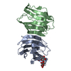
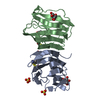
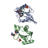
| Entry | Database: PDB / ID: 1gzw | |||||||||
|---|---|---|---|---|---|---|---|---|---|---|
| Title | X-RAY CRYSTAL STRUCTURE OF HUMAN GALECTIN-1 | |||||||||
 Components Components | (GALECTIN-1) x 2 | |||||||||
 Keywords Keywords | LECTIN / CARBOHYDRATE-BINDING PROTEINS / GALACTOSIDES / GALECTIN / CARBOHYDRATE-BINDING SITE | |||||||||
| Function / homology |  Function and homology information Function and homology informationgalectin complex / lactose binding / negative regulation of T-helper 17 cell lineage commitment / myoblast differentiation / plasma cell differentiation / T cell costimulation / laminin binding / Post-translational protein phosphorylation / cell-cell adhesion / Regulation of Insulin-like Growth Factor (IGF) transport and uptake by Insulin-like Growth Factor Binding Proteins (IGFBPs) ...galectin complex / lactose binding / negative regulation of T-helper 17 cell lineage commitment / myoblast differentiation / plasma cell differentiation / T cell costimulation / laminin binding / Post-translational protein phosphorylation / cell-cell adhesion / Regulation of Insulin-like Growth Factor (IGF) transport and uptake by Insulin-like Growth Factor Binding Proteins (IGFBPs) / positive regulation of inflammatory response / extracellular matrix / regulation of apoptotic process / positive regulation of viral entry into host cell / positive regulation of canonical NF-kappaB signal transduction / positive regulation of apoptotic process / endoplasmic reticulum lumen / receptor ligand activity / apoptotic process / : / RNA binding / extracellular exosome / extracellular region / plasma membrane / cytosol / cytoplasm Similarity search - Function | |||||||||
| Biological species |  HOMO SAPIENS (human) HOMO SAPIENS (human) | |||||||||
| Method |  X-RAY DIFFRACTION / SYNCHROTRON / MOLECULAR REPLACEMENT / Resolution: 1.7 Å X-RAY DIFFRACTION / SYNCHROTRON / MOLECULAR REPLACEMENT / Resolution: 1.7 Å | |||||||||
 Authors Authors | Lopez-Lucendo, M.I.F. / Gabius, H.J. / Romero, A. | |||||||||
 Citation Citation | Journal: J.Mol.Biol. / Year: 2004 Title: Growth-Regulatory Human Galectin-1: Crystallographic Characterisation of the Structural Changes Induced by Single-Site Mutations and Their Impact on the Thermodynamics of Ligand Binding Authors: Lopez-Lucendo, M.I.F. / Solis, D. / Andre, S. / Hirabayashi, J. / Kasai, K. / Kaltner, H. / Gabius, H.J. / Romero, A. | |||||||||
| History |
| |||||||||
| Remark 700 | SHEET THE SHEET STRUCTURE OF THIS MOLECULE IS BIFURCATED. IN ORDER TO REPRESENT THIS FEATURE IN ... SHEET THE SHEET STRUCTURE OF THIS MOLECULE IS BIFURCATED. IN ORDER TO REPRESENT THIS FEATURE IN THE SHEET RECORDS BELOW, TWO SHEETS ARE DEFINED. |
- Structure visualization
| Structure viewer | Molecule: MolmilJmol/JSmol |
|---|
- Downloads & links
-Download
| PDBx/mmCIF format | 1gzw.cif.gz | 73.1 KB | Display | PDBx/mmCIF format |
|---|---|---|---|---|
| PDB format | pdb1gzw.ent.gz | 53.2 KB | Display | PDB format |
| PDBx/mmJSON format | 1gzw.json.gz | Tree view | PDBx/mmJSON format | |
| Others |  Other downloads Other downloads |
-Validation report
| Arichive directory | https://data.pdbj.org/pub/pdb/validation_reports/gz/1gzwftp://data.pdbj.org/pub/pdb/validation_reports/gz/1gzw | HTTPS FTP |
|---|
-Related structure data
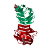
| Related structure data |  1w6mC  1w6nC 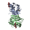 1w6oC 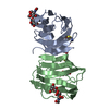 1w6pC  1w6qC 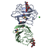 1sltS S: Starting model for refinement C: citing same article ( |
|---|---|
| Similar structure data |


-Links
 PDBj
PDBj


- Assembly
| Deposited unit | 
| ||||||||
|---|---|---|---|---|---|---|---|---|---|
| 1 |
| ||||||||
| Unit cell |
| ||||||||
| Noncrystallographic symmetry (NCS) | NCS oper: (Code: given Matrix: (-0.40139, -0.37095, 0.83743), Vector: |
-Components
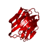
-Protein , 2 types, 2 molecules AB
| #1: Protein | Mass: 14615.477 Da / Num. of mol.: 1 Source method: isolated from a genetically manipulated source Source: (gene. exp.) HOMO SAPIENS (human) / Plasmid: PH14GAL / Production host:  |
|---|---|
| #2: Protein | Mass: 14599.477 Da / Num. of mol.: 1 Source method: isolated from a genetically manipulated source Source: (gene. exp.) HOMO SAPIENS (human) / Plasmid: PH14GAL / Production host: |
-Sugars , 1 types, 2 molecules
| #3: Polysaccharide |   Source method: isolated from a genetically manipulated source Details: oligosaccharide / References: beta-lactose |
|---|
-Non-polymers , 3 types, 290 molecules 


| #4: Chemical | ChemComp-BME /  Mass: 78.133 Da / Num. of mol.: 5 / Source method: obtained synthetically / Formula: C2H6OS Mass: 78.133 Da / Num. of mol.: 5 / Source method: obtained synthetically / Formula: C2H6OS#5: Chemical | ChemComp-SO4 / |  Mass: 96.063 Da / Num. of mol.: 1 / Source method: obtained synthetically / Formula: SO4 Mass: 96.063 Da / Num. of mol.: 1 / Source method: obtained synthetically / Formula: SO4#6: Water | ChemComp-HOH / | Mass: 18.015 Da / Num. of mol.: 284 / Source method: isolated from a natural source / Formula: H2O |
|---|
-Experimental details
-Experiment
| Experiment | Method: X-RAY DIFFRACTION / Number of used crystals: 1 |
|---|
- Sample preparation
| Crystal | Density Matthews: 2.64 Å3/Da / Density % sol: 53.12 % | ||||||||||||||||||||||||||||||||||||
|---|---|---|---|---|---|---|---|---|---|---|---|---|---|---|---|---|---|---|---|---|---|---|---|---|---|---|---|---|---|---|---|---|---|---|---|---|---|
| Crystal grow | Method: vapor diffusion, sitting drop / pH: 5 Details: CRYSTALS WERE OBTAINED IN SITTING DROPS BY MIXING EQUAL VOLUMES OF THE PROTEIN SOLUTION (10 MG/ML) AND THE PRECIPITATING BUFFER (2M AMMONIUM SULPHATE AND 1% BETA-MERCAPTO ETHANOL, PH 5.0) | ||||||||||||||||||||||||||||||||||||
| Crystal grow | *PLUS pH: 7 / Method: vapor diffusion, sitting drop | ||||||||||||||||||||||||||||||||||||
| Components of the solutions | *PLUS
|
-Data collection
| Diffraction | Mean temperature: 100 K |
|---|---|
| Diffraction source | Source: SYNCHROTRON / Site: EMBL/DESY, HAMBURG  / Beamline: X31 / Wavelength: 1.01 / Beamline: X31 / Wavelength: 1.01 |
| Detector | Type: MARRESEARCH / Detector: IMAGE PLATE / Date: Mar 15, 2001 |
| Radiation | Protocol: SINGLE WAVELENGTH / Monochromatic (M) / Laue (L): M / Scattering type: x-ray |
| Radiation wavelength | Wavelength: 1.01 Å / Relative weight: 1 |
| Reflection | Resolution: 1.7→34.41 Å / Num. obs: 34966 / % possible obs: 96.1 % / Redundancy: 3.9 % / Biso Wilson estimate: 16.4 Å2 / Rmerge(I) obs: 0.049 / Net I/σ(I): 12.2 |
| Reflection shell | Resolution: 1.7→1.76 Å / Redundancy: 5.2 % / Rmerge(I) obs: 0.337 / Mean I/σ(I) obs: 5.8 / % possible all: 91.4 |
| Reflection | *PLUS Highest resolution: 1.7 Å / Redundancy: 3.9 % / Num. measured all: 209342 / Rmerge(I) obs: 0.049 |
| Reflection shell | *PLUS % possible obs: 91.4 % / Rmerge(I) obs: 0.337 / Mean I/σ(I) obs: 5.8 |
- Processing
| Software |
| ||||||||||||||||||||||||||||||||||||||||||||||||||||||||||||||||||||||||||||||||
|---|---|---|---|---|---|---|---|---|---|---|---|---|---|---|---|---|---|---|---|---|---|---|---|---|---|---|---|---|---|---|---|---|---|---|---|---|---|---|---|---|---|---|---|---|---|---|---|---|---|---|---|---|---|---|---|---|---|---|---|---|---|---|---|---|---|---|---|---|---|---|---|---|---|---|---|---|---|---|---|---|---|
| Refinement | Method to determine structure: MOLECULAR REPLACEMENT Starting model: PDB ENTRY 1SLT Resolution: 1.7→34.41 Å / Rfactor Rfree error: 0.006 / Data cutoff high absF: 1383157.02 / Isotropic thermal model: RESTRAINED / Cross valid method: THROUGHOUT / σ(F): 0
| ||||||||||||||||||||||||||||||||||||||||||||||||||||||||||||||||||||||||||||||||
| Solvent computation | Solvent model: FLAT MODEL / Bsol: 52.2881 Å2 / ksol: 0.361596 e/Å3 | ||||||||||||||||||||||||||||||||||||||||||||||||||||||||||||||||||||||||||||||||
| Displacement parameters | Biso mean: 23.3 Å2
| ||||||||||||||||||||||||||||||||||||||||||||||||||||||||||||||||||||||||||||||||
| Refine analyze |
| ||||||||||||||||||||||||||||||||||||||||||||||||||||||||||||||||||||||||||||||||
| Refinement step | Cycle: LAST / Resolution: 1.7→34.41 Å
| ||||||||||||||||||||||||||||||||||||||||||||||||||||||||||||||||||||||||||||||||
| Refine LS restraints |
| ||||||||||||||||||||||||||||||||||||||||||||||||||||||||||||||||||||||||||||||||
| LS refinement shell | Resolution: 1.7→1.81 Å / Rfactor Rfree error: 0.021 / Total num. of bins used: 6
| ||||||||||||||||||||||||||||||||||||||||||||||||||||||||||||||||||||||||||||||||
| Xplor file |
| ||||||||||||||||||||||||||||||||||||||||||||||||||||||||||||||||||||||||||||||||
| Refinement | *PLUS Highest resolution: 1.7 Å / Rfactor Rfree: 0.228 / Rfactor Rwork: 0.202 | ||||||||||||||||||||||||||||||||||||||||||||||||||||||||||||||||||||||||||||||||
| Solvent computation | *PLUS | ||||||||||||||||||||||||||||||||||||||||||||||||||||||||||||||||||||||||||||||||
| Displacement parameters | *PLUS | ||||||||||||||||||||||||||||||||||||||||||||||||||||||||||||||||||||||||||||||||
| Refine LS restraints | *PLUS
|
