 Movie
Movie Controller

[English] 日本語
 Yorodumi
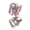
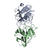
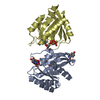
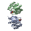
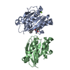
- PDB-6rux: P46, an immunodominant surface protein from Mycoplasma hyopneumoniae -
+ Open data

- Basic information
| Entry | Database: PDB / ID: 6rux | ||||||||||||
|---|---|---|---|---|---|---|---|---|---|---|---|---|---|
| Title | P46, an immunodominant surface protein from Mycoplasma hyopneumoniae | ||||||||||||
 Components Components | 46 kDa surface antigen | ||||||||||||
 Keywords Keywords | PROTEIN BINDING / immunodominant surface protein Mycoplasma hyopneumoniae P46 | ||||||||||||
| Function / homology |  Function and homology information Function and homology informationouter membrane-bounded periplasmic space / carbohydrate binding / plasma membrane Similarity search - Function | ||||||||||||
| Biological species |  Mycoplasma hyopneumoniae J (bacteria) Mycoplasma hyopneumoniae J (bacteria) | ||||||||||||
| Method |  X-RAY DIFFRACTION / SYNCHROTRON / MOLECULAR REPLACEMENT / Resolution: 2.5 Å X-RAY DIFFRACTION / SYNCHROTRON / MOLECULAR REPLACEMENT / Resolution: 2.5 Å | ||||||||||||
 Authors Authors | Guasch, A. / Gonzalez-Gonzalez, L. / Fita, I. | ||||||||||||
| Funding support |  Spain, 2items Spain, 2items
| ||||||||||||
 Citation Citation | Journal: Acta Crystallogr D Struct Biol / Year: 2020 Title: Structure of P46, an immunodominant surface protein from Mycoplasma hyopneumoniae: interaction with a monoclonal antibody. Authors: Guasch, A. / Montane, J. / Moros, A. / Pinol, J. / Sitja, M. / Gonzalez-Gonzalez, L. / Fita, I. | ||||||||||||
| History |
|
- Structure visualization
| Structure viewer | Molecule: MolmilJmol/JSmol |
|---|
- Downloads & links
-Download
| PDBx/mmCIF format | 6rux.cif.gz | 226.5 KB | Display | PDBx/mmCIF format |
|---|---|---|---|---|
| PDB format | pdb6rux.ent.gz | 179.3 KB | Display | PDB format |
| PDBx/mmJSON format | 6rux.json.gz | Tree view | PDBx/mmJSON format | |
| Others |  Other downloads Other downloads |
-Validation report
| Arichive directory | https://data.pdbj.org/pub/pdb/validation_reports/ru/6ruxftp://data.pdbj.org/pub/pdb/validation_reports/ru/6rux | HTTPS FTP |
|---|
-Related structure data
| Related structure data |  6rnnC 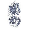 6rqgSC 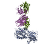 6s3tC S: Starting model for refinement C: citing same article ( |
|---|---|
| Similar structure data |


-Links
 PDBj
PDBj


- Assembly
| Deposited unit | 
| ||||||||||
|---|---|---|---|---|---|---|---|---|---|---|---|
| 1 | 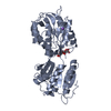
| ||||||||||
| 2 | 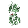
| ||||||||||
| 3 | 
| ||||||||||
| Unit cell |
|
-Components
| #1: Protein | Mass: 42426.508 Da / Num. of mol.: 3 Source method: isolated from a genetically manipulated source Source: (gene. exp.) Mycoplasma hyopneumoniae J (bacteria) / Strain: J / ATCC 25934 / NCTC 10110 / Gene: p46, MHJ_0511Production host: References: UniProt: P0C0J8 #2: Polysaccharide |   Source method: isolated from a genetically manipulated source Details: oligosaccharide / References: alpha-maltose #3: Chemical |   Mass: 22.990 Da / Num. of mol.: 3 / Source method: obtained synthetically / Formula: Na Mass: 22.990 Da / Num. of mol.: 3 / Source method: obtained synthetically / Formula: Na#4: Water | ChemComp-HOH / |  Mass: 18.015 Da / Num. of mol.: 95 / Source method: isolated from a natural source / Formula: H2O Mass: 18.015 Da / Num. of mol.: 95 / Source method: isolated from a natural source / Formula: H2OHas ligand of interest | Y | |
|---|
-Experimental details
-Experiment
| Experiment | Method: X-RAY DIFFRACTION / Number of used crystals: 1 |
|---|
- Sample preparation
| Crystal | Density Matthews: 2.95 Å3/Da / Density % sol: 58.25 % |
|---|---|
| Crystal grow | Temperature: 298 K / Method: vapor diffusion, hanging drop Details: 0.1 M Tris pH 8.5 0.2 M Sodium Acetate 32% PEG 4000 3% Maltose |
-Data collection
| Diffraction | Mean temperature: 100 K / Serial crystal experiment: N |
|---|---|
| Diffraction source | Source: SYNCHROTRON / Site: ALBA / Beamline: XALOC / Wavelength: 0.9793 Å |
| Detector | Type: DECTRIS PILATUS3 6M / Detector: PIXEL / Date: May 17, 2017 |
| Radiation | Protocol: SINGLE WAVELENGTH / Monochromatic (M) / Laue (L): M / Scattering type: x-ray |
| Radiation wavelength | Wavelength: 0.9793 Å / Relative weight: 1 |
| Reflection | Resolution: 2.5→60 Å / Num. obs: 52027 / % possible obs: 60 % / Redundancy: 9.3 % / CC1/2: 0.99 / Rmerge(I) obs: 0.02993 / Net I/σ(I): 35.42 |
| Reflection shell | Resolution: 2.5→2.6 Å / Num. unique obs: 5152 / CC1/2: 0.74 |
- Processing
| Software |
| ||||||||||||||||||||||||
|---|---|---|---|---|---|---|---|---|---|---|---|---|---|---|---|---|---|---|---|---|---|---|---|---|---|
| Refinement | Method to determine structure: MOLECULAR REPLACEMENT Starting model: 6RQG Resolution: 2.5→60 Å / Cross valid method: THROUGHOUT
| ||||||||||||||||||||||||
| Displacement parameters | Biso mean: 75.8 Å2 | ||||||||||||||||||||||||
| Refinement step | Cycle: LAST / Resolution: 2.5→60 Å
| ||||||||||||||||||||||||
| Refine LS restraints |
|
