 Movie
Movie Controller

[English] 日本語
 Yorodumi
- PDB-6rum: Crystal structure of GFP-LAMA-G97 - a GFP enhancer nanobody with ... -
+ Open data

- Basic information
| Entry | Database: PDB / ID: 6rum | ||||||
|---|---|---|---|---|---|---|---|
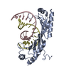
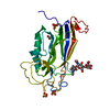
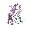
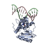
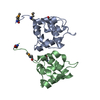
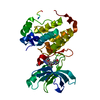
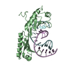
| Title | Crystal structure of GFP-LAMA-G97 - a GFP enhancer nanobody with cpDHFR insertion and TMP and NADPH | ||||||
 Components Components | GFP-LAMA-G97 a GFP enhancer nanobody with cpDHFR insertion | ||||||
 Keywords Keywords | PROTEIN BINDING / LAMA / nanobody / circular permutant of DHFR / TMP / NADPH | ||||||
| Function / homology |  Function and homology information Function and homology informationmethotrexate binding / dihydrofolic acid binding / 10-formyltetrahydrofolate biosynthetic process / response to methotrexate / folic acid biosynthetic process / folic acid binding / NADP+ binding / dihydrofolate metabolic process / dihydrofolate reductase / dihydrofolate reductase activity ...methotrexate binding / dihydrofolic acid binding / 10-formyltetrahydrofolate biosynthetic process / response to methotrexate / folic acid biosynthetic process / folic acid binding / NADP+ binding / dihydrofolate metabolic process / dihydrofolate reductase / dihydrofolate reductase activity / folic acid metabolic process / tetrahydrofolate biosynthetic process / NADPH binding / one-carbon metabolic process / NADP binding / response to xenobiotic stimulus / response to antibiotic / cytosol Similarity search - Function | ||||||
| Biological species |   | ||||||
| Method |  X-RAY DIFFRACTION / SYNCHROTRON / MOLECULAR REPLACEMENT / Resolution: 1.6 Å X-RAY DIFFRACTION / SYNCHROTRON / MOLECULAR REPLACEMENT / Resolution: 1.6 Å | ||||||
 Authors Authors | Farrants, H. / Tarnawski, M. / Mueller, T.G. / Otsuka, S. / Hiblot, J. / Koch, B. / Kueblbeck, M. / Kraeusslich, H.-G. / Ellenberg, J. / Johnsson, K. | ||||||
 Citation Citation | Journal: Nat.Methods / Year: 2020 Title: Chemogenetic Control of Nanobodies. Authors: Farrants, H. / Tarnawski, M. / Muller, T.G. / Otsuka, S. / Hiblot, J. / Koch, B. / Kueblbeck, M. / Krausslich, H.G. / Ellenberg, J. / Johnsson, K. | ||||||
| History |
|
- Structure visualization
| Structure viewer | Molecule: MolmilJmol/JSmol |
|---|
- Downloads & links
-Download
| PDBx/mmCIF format | 6rum.cif.gz | 80.9 KB | Display | PDBx/mmCIF format |
|---|---|---|---|---|
| PDB format | pdb6rum.ent.gz | 56.6 KB | Display | PDB format |
| PDBx/mmJSON format | 6rum.json.gz | Tree view | PDBx/mmJSON format | |
| Others |  Other downloads Other downloads |
-Validation report
| Arichive directory | https://data.pdbj.org/pub/pdb/validation_reports/ru/6rumftp://data.pdbj.org/pub/pdb/validation_reports/ru/6rum | HTTPS FTP |
|---|
-Related structure data
| Related structure data |  6rulC 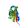 5h8dS  5uiiS C: citing same article ( S: Starting model for refinement |
|---|---|
| Similar structure data |


-Links
 PDBj
PDBj


- Assembly
| Deposited unit | 
| ||||||||
|---|---|---|---|---|---|---|---|---|---|
| 1 |
| ||||||||
| Unit cell |
|
-Components
-Antibody , 1 types, 1 molecules A
| #1: Antibody | Mass: 30984.646 Da / Num. of mol.: 1 Source method: isolated from a genetically manipulated source Source: (gene. exp.) Gene: folA, tmrA, b0048, JW0047 / Production host: |
|---|
-Non-polymers , 6 types, 302 molecules 


| #2: Chemical | ChemComp-TOP /  Mass: 290.318 Da / Num. of mol.: 1 / Source method: obtained synthetically / Formula: C14H18N4O3 / Feature type: SUBJECT OF INVESTIGATION / Comment: antibiotic*YM Mass: 290.318 Da / Num. of mol.: 1 / Source method: obtained synthetically / Formula: C14H18N4O3 / Feature type: SUBJECT OF INVESTIGATION / Comment: antibiotic*YM | ||||||
|---|---|---|---|---|---|---|---|
| #3: Chemical | ChemComp-NDP /  Mass: 745.421 Da / Num. of mol.: 1 / Source method: obtained synthetically / Formula: C21H30N7O17P3 / Feature type: SUBJECT OF INVESTIGATION Mass: 745.421 Da / Num. of mol.: 1 / Source method: obtained synthetically / Formula: C21H30N7O17P3 / Feature type: SUBJECT OF INVESTIGATION | ||||||
| #4: Chemical |  Mass: 35.453 Da / Num. of mol.: 3 / Source method: obtained synthetically / Formula: Cl Mass: 35.453 Da / Num. of mol.: 3 / Source method: obtained synthetically / Formula: Cl#5: Chemical | ChemComp-PG4 / |  Mass: 194.226 Da / Num. of mol.: 1 / Source method: obtained synthetically / Formula: C8H18O5 / Comment: precipitant*YM Mass: 194.226 Da / Num. of mol.: 1 / Source method: obtained synthetically / Formula: C8H18O5 / Comment: precipitant*YM#6: Chemical | ChemComp-PEG / |  Mass: 106.120 Da / Num. of mol.: 1 / Source method: obtained synthetically / Formula: C4H10O3 Mass: 106.120 Da / Num. of mol.: 1 / Source method: obtained synthetically / Formula: C4H10O3#7: Water | ChemComp-HOH / | Mass: 18.015 Da / Num. of mol.: 295 / Source method: isolated from a natural source / Formula: H2O |
-Experimental details
-Experiment
| Experiment | Method: X-RAY DIFFRACTION / Number of used crystals: 1 |
|---|
- Sample preparation
| Crystal | Density Matthews: 2.32 Å3/Da / Density % sol: 47.02 % |
|---|---|
| Crystal grow | Temperature: 293 K / Method: vapor diffusion Details: 0.1 M MES pH 6.0, 20% (w/v) PEG 6000, 1.0 M lithium chloride |
-Data collection
| Diffraction | Mean temperature: 100 K / Serial crystal experiment: N |
|---|---|
| Diffraction source | Source: SYNCHROTRON / Site: SLS  / Beamline: X10SA / Wavelength: 0.99999 Å / Beamline: X10SA / Wavelength: 0.99999 Å |
| Detector | Type: DECTRIS PILATUS 6M / Detector: PIXEL / Date: Apr 19, 2018 |
| Radiation | Monochromator: Si(111) / Protocol: SINGLE WAVELENGTH / Monochromatic (M) / Laue (L): M / Scattering type: x-ray |
| Radiation wavelength | Wavelength: 0.99999 Å / Relative weight: 1 |
| Reflection | Resolution: 1.6→50 Å / Num. obs: 37239 / % possible obs: 99.1 % / Redundancy: 3.3 % / Rmerge(I) obs: 0.054 / Net I/σ(I): 13.23 |
| Reflection shell | Resolution: 1.6→1.7 Å / Redundancy: 3.2 % / Rmerge(I) obs: 0.49 / Mean I/σ(I) obs: 2.43 / Num. unique obs: 6123 / % possible all: 98.6 |
- Processing
| Software |
| ||||||||||||||||||||||||||||||||||||||||||||||||||||||||||||||||||||||||||||||||||||||||||||||||||
|---|---|---|---|---|---|---|---|---|---|---|---|---|---|---|---|---|---|---|---|---|---|---|---|---|---|---|---|---|---|---|---|---|---|---|---|---|---|---|---|---|---|---|---|---|---|---|---|---|---|---|---|---|---|---|---|---|---|---|---|---|---|---|---|---|---|---|---|---|---|---|---|---|---|---|---|---|---|---|---|---|---|---|---|---|---|---|---|---|---|---|---|---|---|---|---|---|---|---|---|
| Refinement | Method to determine structure: MOLECULAR REPLACEMENT Starting model: 5UII, 5H8D Resolution: 1.6→47.715 Å / SU ML: 0.2 / Cross valid method: FREE R-VALUE / σ(F): 1.37 / Phase error: 24.47
| ||||||||||||||||||||||||||||||||||||||||||||||||||||||||||||||||||||||||||||||||||||||||||||||||||
| Solvent computation | Shrinkage radii: 0.9 Å / VDW probe radii: 1.11 Å | ||||||||||||||||||||||||||||||||||||||||||||||||||||||||||||||||||||||||||||||||||||||||||||||||||
| Refinement step | Cycle: LAST / Resolution: 1.6→47.715 Å
| ||||||||||||||||||||||||||||||||||||||||||||||||||||||||||||||||||||||||||||||||||||||||||||||||||
| Refine LS restraints |
| ||||||||||||||||||||||||||||||||||||||||||||||||||||||||||||||||||||||||||||||||||||||||||||||||||
| LS refinement shell |
|
