 Movie
Movie Controller

[English] 日本語
 Yorodumi
- PDB-6rmv: The crystal structure of a TRP channel peptide bound to a G prote... -
+ Open data

- Basic information
| Entry | Database: PDB / ID: 6rmv | |||||||||
|---|---|---|---|---|---|---|---|---|---|---|
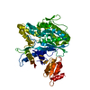
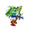
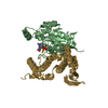
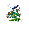
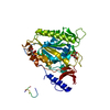
| Title | The crystal structure of a TRP channel peptide bound to a G protein beta gamma heterodimer | |||||||||
 Components Components |
| |||||||||
 Keywords Keywords | PROTEIN BINDING / G protein / TRP Channel / Inhibitor | |||||||||
| Function / homology |  Function and homology information Function and homology informationnew growing cell tip / Activation of the phototransduction cascade / retinal rod cell development / Olfactory Signaling Pathway / Synthesis, secretion, and inactivation of Glucagon-like Peptide-1 (GLP-1) / Sensory perception of sweet, bitter, and umami (glutamate) taste / G beta:gamma signalling through PLC beta / Presynaptic function of Kainate receptors / Prostacyclin signalling through prostacyclin receptor / G alpha (z) signalling events ...new growing cell tip / Activation of the phototransduction cascade / retinal rod cell development / Olfactory Signaling Pathway / Synthesis, secretion, and inactivation of Glucagon-like Peptide-1 (GLP-1) / Sensory perception of sweet, bitter, and umami (glutamate) taste / G beta:gamma signalling through PLC beta / Presynaptic function of Kainate receptors / Prostacyclin signalling through prostacyclin receptor / G alpha (z) signalling events / Glucagon-type ligand receptors / G beta:gamma signalling through PI3Kgamma / G beta:gamma signalling through CDC42 / Adrenaline,noradrenaline inhibits insulin secretion / ADP signalling through P2Y purinoceptor 12 / Cooperation of PDCL (PhLP1) and TRiC/CCT in G-protein beta folding / Thromboxane signalling through TP receptor / G beta:gamma signalling through BTK / Thrombin signalling through proteinase activated receptors (PARs) / Activation of G protein gated Potassium channels / Inhibition of voltage gated Ca2+ channels via Gbeta/gamma subunits / G alpha (s) signalling events / Ca2+ pathway / G-protein activation / Extra-nuclear estrogen signaling / G alpha (12/13) signalling events / G alpha (q) signalling events / Vasopressin regulates renal water homeostasis via Aquaporins / GPER1 signaling / Glucagon-like Peptide-1 (GLP1) regulates insulin secretion / zinc ion transmembrane transporter activity / cellular response to light stimulus / zinc ion transmembrane transport / ADP signalling through P2Y purinoceptor 1 / High laminar flow shear stress activates signaling by PIEZO1 and PECAM1:CDH5:KDR in endothelial cells / cell tip / temperature-gated ion channel activity / G alpha (i) signalling events / TRP channels / metal ion transport / G protein-coupled glutamate receptor signaling pathway / photoreceptor outer segment membrane / spectrin binding / alkylglycerophosphoethanolamine phosphodiesterase activity / sodium ion transport / calcium ion import across plasma membrane / phototransduction, visible light / monoatomic cation transmembrane transport / photoreceptor outer segment / monoatomic cation transport / monoatomic cation channel activity / cardiac muscle cell apoptotic process / phosphatidylinositol-4,5-bisphosphate binding / photoreceptor inner segment / visual perception / protein tetramerization / calcium ion transmembrane transport / calcium channel activity / G-protein beta/gamma-subunit complex binding / intracellular protein localization / myelin sheath / heterotrimeric G-protein complex / G-protein beta-subunit binding / sensory perception of taste / signaling receptor complex adaptor activity / positive regulation of cytosolic calcium ion concentration / retina development in camera-type eye / cell body / GTPase binding / cellular response to hypoxia / protein homotetramerization / phospholipase C-activating G protein-coupled receptor signaling pathway / calmodulin binding / cell population proliferation / G protein-coupled receptor signaling pathway / axon / GTPase activity / synapse / dendrite / endoplasmic reticulum membrane / protein-containing complex binding / signal transduction / endoplasmic reticulum / plasma membrane / cytoplasm Similarity search - Function | |||||||||
| Biological species |  | |||||||||
| Method |  X-RAY DIFFRACTION / SYNCHROTRON / MOLECULAR REPLACEMENT / molecular replacement / Resolution: 1.94 Å X-RAY DIFFRACTION / SYNCHROTRON / MOLECULAR REPLACEMENT / molecular replacement / Resolution: 1.94 Å | |||||||||
 Authors Authors | Gruss, F. / Oberwinkler, J. / Ulens, C. | |||||||||
| Funding support | 2items
| |||||||||
 Citation Citation | Journal: Proc.Natl.Acad.Sci.USA / Year: 2020 Title: The structural basis for an on-off switch controlling G beta gamma-mediated inhibition of TRPM3 channels. Authors: Behrendt, M. / Gruss, F. / Enzeroth, R. / Dembla, S. / Zhao, S. / Crassous, P.A. / Mohr, F. / Nys, M. / Louros, N. / Gallardo, R. / Zorzini, V. / Wagner, D. / Economou, A. / Rousseau, F. / ...Authors: Behrendt, M. / Gruss, F. / Enzeroth, R. / Dembla, S. / Zhao, S. / Crassous, P.A. / Mohr, F. / Nys, M. / Louros, N. / Gallardo, R. / Zorzini, V. / Wagner, D. / Economou, A. / Rousseau, F. / Schymkowitz, J. / Philipp, S.E. / Rohacs, T. / Ulens, C. / Oberwinkler, J. | |||||||||
| History |
|
- Structure visualization
| Structure viewer | Molecule: MolmilJmol/JSmol |
|---|
- Downloads & links
-Download
| PDBx/mmCIF format | 6rmv.cif.gz | 97.1 KB | Display | PDBx/mmCIF format |
|---|---|---|---|---|
| PDB format | pdb6rmv.ent.gz | 71.9 KB | Display | PDB format |
| PDBx/mmJSON format | 6rmv.json.gz | Tree view | PDBx/mmJSON format | |
| Others |  Other downloads Other downloads |
-Validation report
| Arichive directory | https://data.pdbj.org/pub/pdb/validation_reports/rm/6rmvftp://data.pdbj.org/pub/pdb/validation_reports/rm/6rmv | HTTPS FTP |
|---|
-Related structure data
| Related structure data |  1xhmS S: Starting model for refinement |
|---|---|
| Similar structure data |

-Links
 PDBj
PDBj


- Assembly
| Deposited unit | 
| ||||||||
|---|---|---|---|---|---|---|---|---|---|
| 1 |
| ||||||||
| Unit cell |
|
-Components
| #1: Protein | Mass: 37416.930 Da / Num. of mol.: 1 Source method: isolated from a genetically manipulated source Source: (gene. exp.)   Spodoptera frugiperda (fall armyworm) / References: UniProt: P62874 Spodoptera frugiperda (fall armyworm) / References: UniProt: P62874 |
|---|---|
| #2: Protein | Mass: 8046.260 Da / Num. of mol.: 1 / Mutation: C68S Source method: isolated from a genetically manipulated source Source: (gene. exp.) Spodoptera frugiperda (fall armyworm) / References: UniProt: P63213 |
| #3: Protein/peptide | Mass: 1731.109 Da / Num. of mol.: 1 / Source method: obtained synthetically Details: TRPM3 exon 17 - encoded residues including 2 N-terminal and 3 C-terminal flanking residues Source: (synth.) |
| #4: Chemical | ChemComp-GOL /   Mass: 92.094 Da / Num. of mol.: 1 / Source method: obtained synthetically / Formula: C3H8O3 Mass: 92.094 Da / Num. of mol.: 1 / Source method: obtained synthetically / Formula: C3H8O3 |
| #5: Water | ChemComp-HOH /  Mass: 18.015 Da / Num. of mol.: 122 / Source method: isolated from a natural source / Formula: H2O Mass: 18.015 Da / Num. of mol.: 122 / Source method: isolated from a natural source / Formula: H2O |
-Experimental details
-Experiment
| Experiment | Method: X-RAY DIFFRACTION / Number of used crystals: 1 |
|---|
- Sample preparation
| Crystal | Density Matthews: 2.22 Å3/Da / Density % sol: 44.58 % |
|---|---|
| Crystal grow | Temperature: 291.15 K / Method: vapor diffusion, sitting drop / pH: 7.5 Details: 0.1 M HEPES sodium pH 7.5, 15% (w/v) polyethylene glycol 4,000, 7.5% (v/v) isopropanol |
-Data collection
| Diffraction | Mean temperature: 100 K / Serial crystal experiment: N |
|---|---|
| Diffraction source | Source: SYNCHROTRON / Site: ESRF  / Beamline: ID30B / Wavelength: 0.9754 Å / Beamline: ID30B / Wavelength: 0.9754 Å |
| Detector | Type: DECTRIS PILATUS3 6M / Detector: PIXEL / Date: Oct 23, 2017 |
| Radiation | Protocol: SINGLE WAVELENGTH / Monochromatic (M) / Laue (L): M / Scattering type: x-ray |
| Radiation wavelength | Wavelength: 0.9754 Å / Relative weight: 1 |
| Reflection | Resolution: 1.94→42.52 Å / Num. obs: 31659 / % possible obs: 99.3 % / Redundancy: 6.5 % / CC1/2: 0.999 / Rmerge(I) obs: 0.073 / Rpim(I) all: 0.046 / Rrim(I) all: 0.087 / Net I/σ(I): 13.2 |
| Reflection shell | Resolution: 1.94→2.01 Å / Redundancy: 6.7 % / Rmerge(I) obs: 1.28 / Mean I/σ(I) obs: 1.4 / Num. unique obs: 3044 / CC1/2: 0.528 / Rpim(I) all: 0.795 / Rrim(I) all: 1.51 / % possible all: 98.3 |
-Phasing
| Phasing | Method: molecular replacement | |||||||||
|---|---|---|---|---|---|---|---|---|---|---|
| Phasing MR | Model details: Phaser MODE: MR_AUTO
|
- Processing
| Software |
| ||||||||||||||||||||||||||||||||||||||||||||||||||||||||||||
|---|---|---|---|---|---|---|---|---|---|---|---|---|---|---|---|---|---|---|---|---|---|---|---|---|---|---|---|---|---|---|---|---|---|---|---|---|---|---|---|---|---|---|---|---|---|---|---|---|---|---|---|---|---|---|---|---|---|---|---|---|---|
| Refinement | Method to determine structure: MOLECULAR REPLACEMENT Starting model: 1XHM Resolution: 1.94→42.52 Å / Cor.coef. Fo:Fc: 0.968 / Cor.coef. Fo:Fc free: 0.952 / SU B: 4.597 / SU ML: 0.124 / Cross valid method: THROUGHOUT / σ(F): 0 / ESU R: 0.165 / ESU R Free: 0.147 Details: HYDROGENS HAVE BEEN ADDED IN THE RIDING POSITIONS U VALUES : REFINED INDIVIDUALLY
| ||||||||||||||||||||||||||||||||||||||||||||||||||||||||||||
| Solvent computation | Ion probe radii: 0.7 Å / Shrinkage radii: 0.7 Å / VDW probe radii: 1 Å | ||||||||||||||||||||||||||||||||||||||||||||||||||||||||||||
| Displacement parameters | Biso max: 105.39 Å2 / Biso mean: 45.023 Å2 / Biso min: 25.1 Å2
| ||||||||||||||||||||||||||||||||||||||||||||||||||||||||||||
| Refinement step | Cycle: final / Resolution: 1.94→42.52 Å
| ||||||||||||||||||||||||||||||||||||||||||||||||||||||||||||
| Refine LS restraints |
| ||||||||||||||||||||||||||||||||||||||||||||||||||||||||||||
| LS refinement shell | Resolution: 1.94→1.99 Å / Rfactor Rfree error: 0
|
