 Movie
Movie Controller


+ Open data

- Basic information
| Entry | Database: PDB / ID: 6r2q | |||||||||
|---|---|---|---|---|---|---|---|---|---|---|
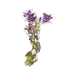
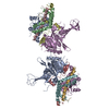
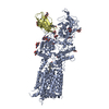
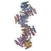
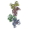
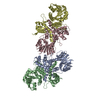
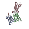
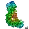
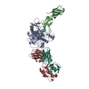
| Title | Structure of the Mtr complex | |||||||||
 Components Components |
| |||||||||
 Keywords Keywords | ELECTRON TRANSPORT / Cytochrome / Membrane protein / greek key / multiheme | |||||||||
| Function / homology |  Function and homology information Function and homology information | |||||||||
| Biological species |  Shewanella baltica (bacteria) Shewanella baltica (bacteria) | |||||||||
| Method |  X-RAY DIFFRACTION / SYNCHROTRON / SAD / Resolution: 2.697 Å X-RAY DIFFRACTION / SYNCHROTRON / SAD / Resolution: 2.697 Å | |||||||||
 Authors Authors | Clarke, T.A. / Edwards, M.J. | |||||||||
| Funding support |  United Kingdom, 2items United Kingdom, 2items
| |||||||||
 Citation Citation | Journal: Cell / Year: 2020 Title: The Crystal Structure of a Biological Insulated Transmembrane Molecular Wire. Authors: Edwards, M.J. / White, G.F. / Butt, J.N. / Richardson, D.J. / Clarke, T.A. | |||||||||
| History |
|
- Structure visualization
| Structure viewer | Molecule: MolmilJmol/JSmol |
|---|
- Downloads & links
-Download
| PDBx/mmCIF format | 6r2q.cif.gz | 583.8 KB | Display | PDBx/mmCIF format |
|---|---|---|---|---|
| PDB format | pdb6r2q.ent.gz | 487.4 KB | Display | PDB format |
| PDBx/mmJSON format | 6r2q.json.gz | Tree view | PDBx/mmJSON format | |
| Others |  Other downloads Other downloads |
-Validation report
| Arichive directory | https://data.pdbj.org/pub/pdb/validation_reports/r2/6r2qftp://data.pdbj.org/pub/pdb/validation_reports/r2/6r2q | HTTPS FTP |
|---|
-Related structure data


-Links
 PDBj
PDBj


- Assembly
| Deposited unit | 
| ||||||||
|---|---|---|---|---|---|---|---|---|---|
| 1 |
| ||||||||
| Unit cell |
|
-Components
-Protein , 3 types, 3 molecules ABC
| #1: Protein | Mass: 36096.930 Da / Num. of mol.: 1 / Source method: isolated from a natural source / Source: (natural) Shewanella baltica (bacteria) / References: UniProt: A0A165K349, UniProt: P0DSN3*PLUS |
|---|---|
| #2: Protein | Mass: 77362.617 Da / Num. of mol.: 1 / Source method: isolated from a natural source / Source: (natural) Shewanella baltica (bacteria) / References: UniProt: A0A165K351, UniProt: P0DSN2*PLUS |
| #3: Protein | Mass: 68131.836 Da / Num. of mol.: 1 / Source method: isolated from a natural source / Source: (natural) Shewanella baltica (bacteria) / References: UniProt: A0A379ZX38, UniProt: P0DSN4*PLUS |
-Non-polymers , 3 types, 46 molecules 


| #4: Chemical | ChemComp-HEC /  Mass: 618.503 Da / Num. of mol.: 20 / Source method: obtained synthetically / Formula: C34H34FeN4O4 Mass: 618.503 Da / Num. of mol.: 20 / Source method: obtained synthetically / Formula: C34H34FeN4O4#5: Chemical | ChemComp-CA /  Mass: 40.078 Da / Num. of mol.: 4 / Source method: obtained synthetically / Formula: Ca Mass: 40.078 Da / Num. of mol.: 4 / Source method: obtained synthetically / Formula: Ca#6: Water | ChemComp-HOH / | Mass: 18.015 Da / Num. of mol.: 22 / Source method: isolated from a natural source / Formula: H2O |
|---|
-Details
| Has protein modification | Y |
|---|
-Experimental details
-Experiment
| Experiment | Method: X-RAY DIFFRACTION / Number of used crystals: 1 |
|---|
- Sample preparation
| Crystal | Density Matthews: 3.71 Å3/Da / Density % sol: 66.82 % |
|---|---|
| Crystal grow | Temperature: 289 K / Method: vapor diffusion, sitting drop Details: 0.1 M Bis-Tris, 0.01 M LDAO, 0.4 M Calcium chloride, 40 % MPD |
-Data collection
| Diffraction | Mean temperature: 100 K / Serial crystal experiment: N | ||||||||||||||||||||||||
|---|---|---|---|---|---|---|---|---|---|---|---|---|---|---|---|---|---|---|---|---|---|---|---|---|---|
| Diffraction source | Source: SYNCHROTRON / Site: Diamond / Beamline: I03 / Wavelength: 0.97624 Å | ||||||||||||||||||||||||
| Detector | Type: DECTRIS PILATUS 300K / Detector: PIXEL / Date: Sep 10, 2018 | ||||||||||||||||||||||||
| Radiation | Protocol: SINGLE WAVELENGTH / Monochromatic (M) / Laue (L): M / Scattering type: x-ray | ||||||||||||||||||||||||
| Radiation wavelength | Wavelength: 0.97624 Å / Relative weight: 1 | ||||||||||||||||||||||||
| Reflection | Entry-ID: 6R2Q / Diffraction-ID: 1
| ||||||||||||||||||||||||
| Reflection shell | Resolution: 2.697→2.814 Å / Redundancy: 4.7 % / Mean I/σ(I) obs: 2.6 / Num. unique obs: 2489 / CC1/2: 0.908 / % possible all: 97.1 |
-Phasing
| Phasing | Method: SAD |
|---|
- Processing
| Software |
| |||||||||||||||||||||||||||||||||||||||||||||||||||||||||||||||||||||||||||||||||||||||||||||||||||||||||||||||||||||||||||||||||||||
|---|---|---|---|---|---|---|---|---|---|---|---|---|---|---|---|---|---|---|---|---|---|---|---|---|---|---|---|---|---|---|---|---|---|---|---|---|---|---|---|---|---|---|---|---|---|---|---|---|---|---|---|---|---|---|---|---|---|---|---|---|---|---|---|---|---|---|---|---|---|---|---|---|---|---|---|---|---|---|---|---|---|---|---|---|---|---|---|---|---|---|---|---|---|---|---|---|---|---|---|---|---|---|---|---|---|---|---|---|---|---|---|---|---|---|---|---|---|---|---|---|---|---|---|---|---|---|---|---|---|---|---|---|---|---|
| Refinement | Method to determine structure: SAD / Resolution: 2.697→73.25 Å / SU ML: 0.36 / Cross valid method: THROUGHOUT / σ(F): 1.37 / Phase error: 30.23
| |||||||||||||||||||||||||||||||||||||||||||||||||||||||||||||||||||||||||||||||||||||||||||||||||||||||||||||||||||||||||||||||||||||
| Solvent computation | Shrinkage radii: 0.9 Å / VDW probe radii: 1.11 Å | |||||||||||||||||||||||||||||||||||||||||||||||||||||||||||||||||||||||||||||||||||||||||||||||||||||||||||||||||||||||||||||||||||||
| Displacement parameters | Biso max: 220.71 Å2 / Biso mean: 62.5529 Å2 / Biso min: 9.31 Å2 | |||||||||||||||||||||||||||||||||||||||||||||||||||||||||||||||||||||||||||||||||||||||||||||||||||||||||||||||||||||||||||||||||||||
| Refinement step | Cycle: final / Resolution: 2.697→73.25 Å
| |||||||||||||||||||||||||||||||||||||||||||||||||||||||||||||||||||||||||||||||||||||||||||||||||||||||||||||||||||||||||||||||||||||
| LS refinement shell | Refine-ID: X-RAY DIFFRACTION / Rfactor Rfree error: 0 / Total num. of bins used: 18
|
