Movie
Movie Controller

[English] 日本語
 Yorodumi
- PDB-6qvd: CryoEM structure of the human ClC-1 chloride channel, CBS state 2 -
+ Open data

- Basic information
| Entry | Database: PDB / ID: 6qvd | ||||||
|---|---|---|---|---|---|---|---|
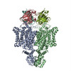
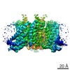
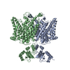
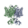
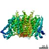
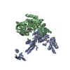
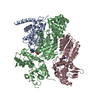
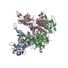
| Title | CryoEM structure of the human ClC-1 chloride channel, CBS state 2 | ||||||
 Components Components | Chloride channel protein 1 | ||||||
 Keywords Keywords | MEMBRANE PROTEIN / chloride channel / CLC1 / CLCN1 | ||||||
| Function / homology |  Function and homology information Function and homology informationneuronal action potential propagation / voltage-gated chloride channel activity / chloride transport / chloride channel complex / muscle contraction / T-tubule / chloride transmembrane transport / Stimuli-sensing channels / protein homodimerization activity / plasma membrane Similarity search - Function | ||||||
| Biological species |  Homo sapiens (human) Homo sapiens (human) | ||||||
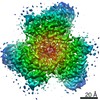
| Method | ELECTRON MICROSCOPY / single particle reconstruction / cryo EM / Resolution: 4.34 Å | ||||||
 Authors Authors | Wang, K.T. / Gourdon, P.E. / Zhou, Z.H. | ||||||
| Funding support |  Denmark, 1items Denmark, 1items
| ||||||
 Citation Citation | Journal: PLoS Biol / Year: 2019 Title: Structure of the human ClC-1 chloride channel. Authors: Kaituo Wang / Sarah Spruce Preisler / Liying Zhang / Yanxiang Cui / Julie Winkel Missel / Christina Grønberg / Kamil Gotfryd / Erik Lindahl / Magnus Andersson / Kirstine Calloe / Pascal F ...Authors: Kaituo Wang / Sarah Spruce Preisler / Liying Zhang / Yanxiang Cui / Julie Winkel Missel / Christina Grønberg / Kamil Gotfryd / Erik Lindahl / Magnus Andersson / Kirstine Calloe / Pascal F Egea / Dan Arne Klaerke / Michael Pusch / Per Amstrup Pedersen / Z Hong Zhou / Pontus Gourdon /    Abstract: ClC-1 protein channels facilitate rapid passage of chloride ions across cellular membranes, thereby orchestrating skeletal muscle excitability. Malfunction of ClC-1 is associated with myotonia ...ClC-1 protein channels facilitate rapid passage of chloride ions across cellular membranes, thereby orchestrating skeletal muscle excitability. Malfunction of ClC-1 is associated with myotonia congenita, a disease impairing muscle relaxation. Here, we present the cryo-electron microscopy (cryo-EM) structure of human ClC-1, uncovering an architecture reminiscent of that of bovine ClC-K and CLC transporters. The chloride conducting pathway exhibits distinct features, including a central glutamate residue ("fast gate") known to confer voltage-dependence (a mechanistic feature not present in ClC-K), linked to a somewhat rearranged central tyrosine and a narrower aperture of the pore toward the extracellular vestibule. These characteristics agree with the lower chloride flux of ClC-1 compared with ClC-K and enable us to propose a model for chloride passage in voltage-dependent CLC channels. Comparison of structures derived from protein studied in different experimental conditions supports the notion that pH and adenine nucleotides regulate ClC-1 through interactions between the so-called cystathionine-β-synthase (CBS) domains and the intracellular vestibule ("slow gating"). The structure also provides a framework for analysis of mutations causing myotonia congenita and reveals a striking correlation between mutated residues and the phenotypic effect on voltage gating, opening avenues for rational design of therapies against ClC-1-related diseases. | ||||||
| History |
|
- Structure visualization
| Movie |
Movie viewer |
|---|---|
| Structure viewer | Molecule: MolmilJmol/JSmol |
- Downloads & links
-Download
| PDBx/mmCIF format | 6qvd.cif.gz | 224.4 KB | Display | PDBx/mmCIF format |
|---|---|---|---|---|
| PDB format | pdb6qvd.ent.gz | 172.5 KB | Display | PDB format |
| PDBx/mmJSON format | 6qvd.json.gz | Tree view | PDBx/mmJSON format | |
| Others |  Other downloads Other downloads |
-Validation report
| Arichive directory | https://data.pdbj.org/pub/pdb/validation_reports/qv/6qvdftp://data.pdbj.org/pub/pdb/validation_reports/qv/6qvd | HTTPS FTP |
|---|
-Related structure data
| Related structure data | 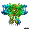 4649MC 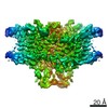 4645C 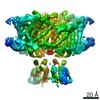 4646C 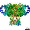 4647C 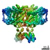 4657C 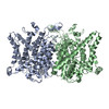 6qv6C  6qvbC 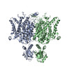 6qvcC 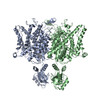 6qvuC C: citing same article ( M: map data used to model this data |
|---|---|
| Similar structure data |
-Links
 PDBj
PDBj
- Assembly
| Deposited unit | 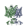
|
|---|---|
| 1 |
|
-Components
| #1: Protein | Mass: 108733.172 Da / Num. of mol.: 2 Source method: isolated from a genetically manipulated source Source: (gene. exp.) Homo sapiens (human) / Gene: CLCN1, CLC1 / Production host:  |
|---|
-Experimental details
-Experiment
| Experiment | Method: ELECTRON MICROSCOPY |
|---|---|
| EM experiment | Aggregation state: PARTICLE / 3D reconstruction method: single particle reconstruction |
- Sample preparation
| Component | Name: human Chloride Channel protein 1 / Type: COMPLEX / Entity ID: all / Source: RECOMBINANT |
|---|---|
| Molecular weight | Value: 0.109 MDa / Experimental value: NO |
| Source (natural) | Organism: Homo sapiens (human) |
| Source (recombinant) | Organism: |
| Buffer solution | pH: 7.5 / Details: 20mM Tris ph7.5 100mM NaCl 0.2mM TCEP |
| Buffer component | Conc.: 20 mM / Name: Tris |
| Specimen | Conc.: 0.6 mg/ml / Embedding applied: NO / Shadowing applied: NO / Staining applied: NO / Vitrification applied: YES |
| Vitrification | Instrument: FEI VITROBOT MARK IV / Cryogen name: ETHANE / Humidity: 100 % / Chamber temperature: 277 K |
- Electron microscopy imaging
| Experimental equipment |  Model: Titan Krios / Image courtesy: FEI Company |
|---|---|
| Microscopy | Model: FEI TITAN KRIOS |
| Electron gun | Electron source:  FIELD EMISSION GUN / Accelerating voltage: 300 kV / Illumination mode: OTHER FIELD EMISSION GUN / Accelerating voltage: 300 kV / Illumination mode: OTHER |
| Electron lens | Mode: BRIGHT FIELD / Cs: 2.7 mm / C2 aperture diameter: 70 µm / Alignment procedure: COMA FREE |
| Specimen holder | Cryogen: NITROGEN / Specimen holder model: FEI TITAN KRIOS AUTOGRID HOLDER |
| Image recording | Average exposure time: 9 sec. / Electron dose: 45 e/Å2 / Detector mode: SUPER-RESOLUTION / Film or detector model: GATAN K2 SUMMIT (4k x 4k) |
| EM imaging optics | Energyfilter slit width: 20 eV |
| Image scans | Movie frames/image: 60 |
- Processing
| EM software |
| ||||||||||||||||||||||||||||||||||||
|---|---|---|---|---|---|---|---|---|---|---|---|---|---|---|---|---|---|---|---|---|---|---|---|---|---|---|---|---|---|---|---|---|---|---|---|---|---|
| CTF correction | Type: PHASE FLIPPING ONLY | ||||||||||||||||||||||||||||||||||||
| Particle selection | Num. of particles selected: 477729 | ||||||||||||||||||||||||||||||||||||
| Symmetry | Point symmetry: C2 (2 fold cyclic) | ||||||||||||||||||||||||||||||||||||
| 3D reconstruction | Resolution: 4.34 Å / Resolution method: FSC 0.143 CUT-OFF / Num. of particles: 74048 / Algorithm: BACK PROJECTION / Num. of class averages: 1 / Symmetry type: POINT | ||||||||||||||||||||||||||||||||||||
| Atomic model building | Protocol: BACKBONE TRACE / Space: REAL | ||||||||||||||||||||||||||||||||||||
| Atomic model building | PDB-ID: 5TQQ Accession code: 5TQQ / Source name: PDB / Type: experimental model |