 Movie
Movie Controller


+ Open data

- Basic information
| Entry | Database: PDB / ID: 3qo6 | ||||||
|---|---|---|---|---|---|---|---|
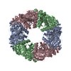
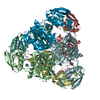
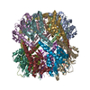
| Title | Crystal structure analysis of the plant protease Deg1 | ||||||
 Components Components |
| ||||||
 Keywords Keywords | PHOTOSYNTHESIS / protease / HtrA / pH-sensor / hydrolase | ||||||
| Function / homology |  Function and homology information Function and homology informationphotosystem II repair / thylakoid lumen / chloroplast thylakoid / chloroplast thylakoid lumen / thylakoid / chloroplast thylakoid membrane / Hydrolases; Acting on peptide bonds (peptidases); Serine endopeptidases / serine-type peptidase activity / protein catabolic process / chloroplast ...photosystem II repair / thylakoid lumen / chloroplast thylakoid / chloroplast thylakoid lumen / thylakoid / chloroplast thylakoid membrane / Hydrolases; Acting on peptide bonds (peptidases); Serine endopeptidases / serine-type peptidase activity / protein catabolic process / chloroplast / serine-type endopeptidase activity / proteolysis / identical protein binding / nucleus / cytosol Similarity search - Function | ||||||
| Biological species |  unidentified (others) | ||||||
| Method |  X-RAY DIFFRACTION / SYNCHROTRON / MOLECULAR REPLACEMENT / molecular replacement / Resolution: 2.5 Å X-RAY DIFFRACTION / SYNCHROTRON / MOLECULAR REPLACEMENT / molecular replacement / Resolution: 2.5 Å | ||||||
 Authors Authors | Clausen, T. | ||||||
 Citation Citation | Journal: Nat.Struct.Mol.Biol. / Year: 2011 Title: Structural adaptation of the plant protease Deg1 to repair photosystem II during light exposure. Authors: Kley, J. / Schmidt, B. / Boyanov, B. / Stolt-Bergner, P.C. / Kirk, R. / Ehrmann, M. / Knopf, R.R. / Naveh, L. / Adam, Z. / Clausen, T. | ||||||
| History |
| ||||||
| Remark 999 | AUTHOR STATES THAT THE PEPTIDES WERE CO-PURIFIED AND CO-CRYSTALLIZED WITH THE DEG1 PROTEIN FROM E. ...AUTHOR STATES THAT THE PEPTIDES WERE CO-PURIFIED AND CO-CRYSTALLIZED WITH THE DEG1 PROTEIN FROM E.COLI, HOWEVER THEIR PRECISE ORIGIN IS UNKNOWN. THEY ARE MODELED AS ALA IN THE DEPOSITED PDB FILE AND HAVE BEEN CHANGED TO UNK DURING PROCESSING. |
- Structure visualization
| Structure viewer | Molecule: MolmilJmol/JSmol |
|---|
- Downloads & links
-Download
| PDBx/mmCIF format | 3qo6.cif.gz | 196.4 KB | Display | PDBx/mmCIF format |
|---|---|---|---|---|
| PDB format | pdb3qo6.ent.gz | 158.5 KB | Display | PDB format |
| PDBx/mmJSON format | 3qo6.json.gz | Tree view | PDBx/mmJSON format | |
| Others |  Other downloads Other downloads |
-Validation report
| Arichive directory | https://data.pdbj.org/pub/pdb/validation_reports/qo/3qo6ftp://data.pdbj.org/pub/pdb/validation_reports/qo/3qo6 | HTTPS FTP |
|---|
-Related structure data
| Related structure data |  3mh7S S: Starting model for refinement |
|---|---|
| Similar structure data |


-Links
 PDBj
PDBj
- Assembly
| Deposited unit | 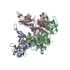
| ||||||||
|---|---|---|---|---|---|---|---|---|---|
| 1 | 
| ||||||||
| Unit cell |
|
-Components
-Protein/peptide , 4 types, 6 molecules DEHIFG
| #2: Protein/peptide | Mass: 613.749 Da / Num. of mol.: 1 Source method: isolated from a genetically manipulated source Source: (gene. exp.) unidentified (others) / Production host:  | ||||
|---|---|---|---|---|---|
| #3: Protein/peptide | Mass: 358.434 Da / Num. of mol.: 3 Source method: isolated from a genetically manipulated source Source: (gene. exp.) unidentified (others) / Production host: #4: Protein/peptide | | Mass: 443.539 Da / Num. of mol.: 1 Source method: isolated from a genetically manipulated source Source: (gene. exp.) unidentified (others) / Production host: #5: Protein/peptide | | Mass: 273.330 Da / Num. of mol.: 1 Source method: isolated from a genetically manipulated source Source: (gene. exp.) unidentified (others) / Production host: |
-Protein / Non-polymers , 2 types, 141 molecules ABC
| #1: Protein | Mass: 36785.508 Da / Num. of mol.: 3 Source method: isolated from a genetically manipulated source Source: (gene. exp.) References: UniProt: O22609, Hydrolases; Acting on peptide bonds (peptidases); Serine endopeptidases #6: Water | ChemComp-HOH / | Mass: 18.015 Da / Num. of mol.: 138 / Source method: isolated from a natural source / Formula: H2O |
|---|
-Details
| Has protein modification | Y |
|---|
-Experimental details
-Experiment
| Experiment | Method: X-RAY DIFFRACTION / Number of used crystals: 1 |
|---|
- Sample preparation
| Crystal | Density Matthews: 3.52 Å3/Da / Density % sol: 65.07 % |
|---|---|
| Crystal grow | Temperature: 292 K / Method: vapor diffusion / pH: 5.2 Details: sodium citrate, ammonium sulfate, lithium sulfate, pH 5.2, vapor diffusion, temperature 292K |
-Data collection
| Diffraction | Mean temperature: 100 K | ||||||||||||||||||||||||||||||||||||||||||||||||||||||||||||||||||
|---|---|---|---|---|---|---|---|---|---|---|---|---|---|---|---|---|---|---|---|---|---|---|---|---|---|---|---|---|---|---|---|---|---|---|---|---|---|---|---|---|---|---|---|---|---|---|---|---|---|---|---|---|---|---|---|---|---|---|---|---|---|---|---|---|---|---|---|
| Diffraction source | Source: SYNCHROTRON / Site: ESRF  / Beamline: ID14-4 / Wavelength: 0.9792 Å / Beamline: ID14-4 / Wavelength: 0.9792 Å | ||||||||||||||||||||||||||||||||||||||||||||||||||||||||||||||||||
| Detector | Type: ADSC QUANTUM 315 / Detector: CCD / Date: Mar 15, 2010 | ||||||||||||||||||||||||||||||||||||||||||||||||||||||||||||||||||
| Radiation | Monochromator: MIRRORS / Protocol: SINGLE WAVELENGTH / Scattering type: x-ray | ||||||||||||||||||||||||||||||||||||||||||||||||||||||||||||||||||
| Radiation wavelength | Wavelength: 0.9792 Å / Relative weight: 1 | ||||||||||||||||||||||||||||||||||||||||||||||||||||||||||||||||||
| Reflection | Resolution: 2.5→20 Å / Num. all: 57809 / Num. obs: 53034 / % possible obs: 98.1 % / Observed criterion σ(F): -1000 / Observed criterion σ(I): -1000 / Rmerge(I) obs: 0.054 / Χ2: 1.083 / Net I/σ(I): 10.7 | ||||||||||||||||||||||||||||||||||||||||||||||||||||||||||||||||||
| Reflection shell |
|
-Phasing
| Phasing | Method: molecular replacement |
|---|
- Processing
| Software |
| ||||||||||||||||||||||||||||
|---|---|---|---|---|---|---|---|---|---|---|---|---|---|---|---|---|---|---|---|---|---|---|---|---|---|---|---|---|---|
| Refinement | Method to determine structure: MOLECULAR REPLACEMENT Starting model: 3MH7 Resolution: 2.5→15 Å / Occupancy max: 1 / Occupancy min: 0.5 / σ(F): 0 / σ(I): 0 / Stereochemistry target values: Engh & Huber
| ||||||||||||||||||||||||||||
| Solvent computation | Bsol: 75.6883 Å2 | ||||||||||||||||||||||||||||
| Displacement parameters | Biso max: 154.97 Å2 / Biso mean: 85.1888 Å2 / Biso min: 34.27 Å2
| ||||||||||||||||||||||||||||
| Refinement step | Cycle: LAST / Resolution: 2.5→15 Å
| ||||||||||||||||||||||||||||
| Refine LS restraints |
| ||||||||||||||||||||||||||||
| Xplor file |
|
