 Movie
Movie Controller

[English] 日本語
 Yorodumi
- PDB-3mh7: HtrA proteases are activated by a conserved mechanism that can be... -
+ Open data

- Basic information
| Entry | Database: PDB / ID: 3mh7 | ||||||
|---|---|---|---|---|---|---|---|
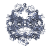
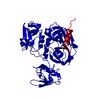
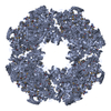
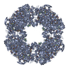
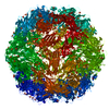
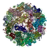
| Title | HtrA proteases are activated by a conserved mechanism that can be triggered by distinct molecular cues | ||||||
 Components Components |
| ||||||
 Keywords Keywords | HYDROLASE / DegP / HtrA / protease / outer membrane protein | ||||||
| Function / homology |  Function and homology information Function and homology informationpeptidase Do / response to temperature stimulus / protein quality control for misfolded or incompletely synthesized proteins / Secretion of toxins / serine-type peptidase activity / peptidase activity / outer membrane-bounded periplasmic space / response to heat / protein folding / response to oxidative stress ...peptidase Do / response to temperature stimulus / protein quality control for misfolded or incompletely synthesized proteins / Secretion of toxins / serine-type peptidase activity / peptidase activity / outer membrane-bounded periplasmic space / response to heat / protein folding / response to oxidative stress / periplasmic space / serine-type endopeptidase activity / proteolysis / identical protein binding / plasma membrane Similarity search - Function | ||||||
| Biological species |  | ||||||
| Method |  X-RAY DIFFRACTION / SYNCHROTRON / SAD / Resolution: 2.961 Å X-RAY DIFFRACTION / SYNCHROTRON / SAD / Resolution: 2.961 Å | ||||||
 Authors Authors | Krojer, T. / Sawa, J. / Huber, R. / Clausen, T. | ||||||
 Citation Citation | Journal: Nat.Struct.Mol.Biol. / Year: 2010 Title: HtrA proteases have a conserved activation mechanism that can be triggered by distinct molecular cues Authors: Krojer, T. / Sawa, J. / Huber, R. / Clausen, T. | ||||||
| History |
|
- Structure visualization
| Structure viewer | Molecule: MolmilJmol/JSmol |
|---|
- Downloads & links
-Download
| PDBx/mmCIF format | 3mh7.cif.gz | 85.9 KB | Display | PDBx/mmCIF format |
|---|---|---|---|---|
| PDB format | pdb3mh7.ent.gz | 67.2 KB | Display | PDB format |
| PDBx/mmJSON format | 3mh7.json.gz | Tree view | PDBx/mmJSON format | |
| Others |  Other downloads Other downloads |
-Validation report
| Arichive directory | https://data.pdbj.org/pub/pdb/validation_reports/mh/3mh7ftp://data.pdbj.org/pub/pdb/validation_reports/mh/3mh7 | HTTPS FTP |
|---|
-Related structure data


-Links
 PDBj
PDBj- Assembly
| Deposited unit | 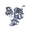
| ||||||||
|---|---|---|---|---|---|---|---|---|---|
| 1 | x 24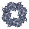
| ||||||||
| Unit cell |
|
-Components
| #1: Protein | Mass: 48582.594 Da / Num. of mol.: 1 / Mutation: S210A Source method: isolated from a genetically manipulated source Source: (gene. exp.) References: UniProt: P0C0V0, Hydrolases; Acting on peptide bonds (peptidases); Serine endopeptidases | ||||
|---|---|---|---|---|---|
| #2: Protein/peptide | Mass: 443.539 Da / Num. of mol.: 2 Source method: isolated from a genetically manipulated source Details: this peptide apparently picked up during protein purification. Source: (gene. exp.) Has protein modification | Y | Sequence details | THE DEPOSITORS COULD NOT DETERMINE THE SEQUENCE FOR CHAIN B, C AND COULD NOT PROVE WHETHER CHIAN B ...THE DEPOSITORS | |
-Experimental details
-Experiment
| Experiment | Method: X-RAY DIFFRACTION / Number of used crystals: 1 |
|---|
- Sample preparation
| Crystal | Density Matthews: 3.45 Å3/Da / Density % sol: 64.32 % |
|---|---|
| Crystal grow | Temperature: 292 K / Method: vapor diffusion / pH: 8.5 Details: PEG 550 MME, NaCl, pH 8.5, VAPOR DIFFUSION, temperature 292K |
-Data collection
| Diffraction | Mean temperature: 100 K |
|---|---|
| Diffraction source | Source: SYNCHROTRON / Site: ESRF  / Beamline: ID23-1 / Beamline: ID23-1 |
| Detector | Type: ADSC QUANTUM 4 / Detector: CCD / Date: Dec 15, 2006 |
| Radiation | Protocol: SINGLE WAVELENGTH / Monochromatic (M) / Laue (L): M / Scattering type: x-ray |
| Radiation wavelength | Relative weight: 1 |
| Reflection | Resolution: 2.961→30 Å / Num. obs: 15145 / % possible obs: 99.7 % / Observed criterion σ(F): -3 / Observed criterion σ(I): -3 / Redundancy: 5.1 % / Biso Wilson estimate: 60.3 Å2 / Rmerge(I) obs: 0.093 / Rsym value: 0.093 / Net I/σ(I): 13 |
| Reflection shell | Resolution: 2.961→3.19 Å / Redundancy: 5.1 % / Rmerge(I) obs: 0.579 / Mean I/σ(I) obs: 2.9 / Rsym value: 0.579 / % possible all: 99.9 |
- Processing
| Software |
| ||||||||||||||||||||||||||||||
|---|---|---|---|---|---|---|---|---|---|---|---|---|---|---|---|---|---|---|---|---|---|---|---|---|---|---|---|---|---|---|---|
| Refinement | Method to determine structure: SAD / Resolution: 2.961→29.321 Å / Occupancy max: 1 / Occupancy min: 1 / FOM work R set: 0.828 / SU ML: 0.37 / Cross valid method: THROUGHOUT / σ(F): 1.33 / Phase error: 23.12 / Stereochemistry target values: ML
| ||||||||||||||||||||||||||||||
| Solvent computation | Shrinkage radii: 0.8 Å / VDW probe radii: 1 Å / Solvent model: FLAT BULK SOLVENT MODEL / Bsol: 53.861 Å2 / ksol: 0.335 e/Å3 | ||||||||||||||||||||||||||||||
| Displacement parameters | Biso max: 176.98 Å2 / Biso mean: 75.679 Å2 / Biso min: 33.13 Å2
| ||||||||||||||||||||||||||||||
| Refinement step | Cycle: LAST / Resolution: 2.961→29.321 Å
| ||||||||||||||||||||||||||||||
| Refine LS restraints |
| ||||||||||||||||||||||||||||||
| LS refinement shell | Refine-ID: X-RAY DIFFRACTION / Total num. of bins used: 5 / % reflection obs: 100 %
|
