 Movie
Movie Controller


+ Open data

- Basic information
| Entry | Database: PDB / ID: 3ou0 | ||||||
|---|---|---|---|---|---|---|---|
| Title | re-refined 3CS0 | ||||||
 Components Components |
| ||||||
 Keywords Keywords | HYDROLASE / protease | ||||||
| Function / homology |  Function and homology information Function and homology informationpeptidase Do / response to temperature stimulus / Secretion of toxins / protein quality control for misfolded or incompletely synthesized proteins / serine-type peptidase activity / peptidase activity / outer membrane-bounded periplasmic space / response to heat / protein folding / response to oxidative stress ...peptidase Do / response to temperature stimulus / Secretion of toxins / protein quality control for misfolded or incompletely synthesized proteins / serine-type peptidase activity / peptidase activity / outer membrane-bounded periplasmic space / response to heat / protein folding / response to oxidative stress / periplasmic space / serine-type endopeptidase activity / proteolysis / identical protein binding / plasma membrane Similarity search - Function | ||||||
| Biological species |  | ||||||
| Method |  X-RAY DIFFRACTION / SAD / Resolution: 3 Å X-RAY DIFFRACTION / SAD / Resolution: 3 Å | ||||||
 Authors Authors | Sauer, R.T. / Grant, R.A. / Kim, S. | ||||||
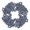
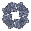
 Citation Citation | Journal: Nature / Year: 2008 Title: Structural basis for the regulated protease and chaperone function of DegP. Authors: Tobias Krojer / Justyna Sawa / Eva Schäfer / Helen R Saibil / Michael Ehrmann / Tim Clausen /  Abstract: All organisms have to monitor the folding state of cellular proteins precisely. The heat-shock protein DegP is a protein quality control factor in the bacterial envelope that is involved in ...All organisms have to monitor the folding state of cellular proteins precisely. The heat-shock protein DegP is a protein quality control factor in the bacterial envelope that is involved in eliminating misfolded proteins and in the biogenesis of outer-membrane proteins. Here we describe the molecular mechanisms underlying the regulated protease and chaperone function of DegP from Escherichia coli. We show that binding of misfolded proteins transforms hexameric DegP into large, catalytically active 12-meric and 24-meric multimers. A structural analysis of these particles revealed that DegP represents a protein packaging device whose central compartment is adaptable to the size and concentration of substrate. Moreover, the inner cavity serves antagonistic functions. Whereas the encapsulation of folded protomers of outer-membrane proteins is protective and might allow safe transit through the periplasm, misfolded proteins are eliminated in the molecular reaction chamber. Oligomer reassembly and concomitant activation on substrate binding may also be critical in regulating other HtrA proteases implicated in protein-folding diseases. | ||||||
| History |
| ||||||
| Remark 0 | THIS ENTRY 3OU0 REFLECTS AN ALTERNATIVE MODELING OF THE ORIGINAL STRUCTURAL DATA R3CS0SF DETERMINED ...THIS ENTRY 3OU0 REFLECTS AN ALTERNATIVE MODELING OF THE ORIGINAL STRUCTURAL DATA R3CS0SF DETERMINED BY AUTHORS OF THE PDB ENTRY 3CS0: T.KROJER,J.SAWA,E.SCHAEFER,H.R.SAIBIL,M.EHRMANN,T.CLAUSEN | ||||||
| Remark 200 | AUTHOR USED THE SF(MR) DATA FROM ENTRY 3CS0 | ||||||
| Remark 999 | THIS ENTRY 3OU0 REFLECTS AN ALTERNATIVE MODELING OF X-RAY DATA R3CS0SF. 3OU0 HAS AN EXTRA CHAIN C ...THIS ENTRY 3OU0 REFLECTS AN ALTERNATIVE MODELING OF X-RAY DATA R3CS0SF. 3OU0 HAS AN EXTRA CHAIN C COMPARING THE ORIGINAL DEPOSITION 3CS0. |
- Structure visualization
| Structure viewer | Molecule: MolmilJmol/JSmol |
|---|
- Downloads & links
-Download
| PDBx/mmCIF format | 3ou0.cif.gz | 85.6 KB | Display | PDBx/mmCIF format |
|---|---|---|---|---|
| PDB format | pdb3ou0.ent.gz | 66.5 KB | Display | PDB format |
| PDBx/mmJSON format | 3ou0.json.gz | Tree view | PDBx/mmJSON format | |
| Others |  Other downloads Other downloads |
-Validation report
| Arichive directory | https://data.pdbj.org/pub/pdb/validation_reports/ou/3ou0ftp://data.pdbj.org/pub/pdb/validation_reports/ou/3ou0 | HTTPS FTP |
|---|
-Related structure data


-Links
 PDBj
PDBj- Assembly
| Deposited unit | 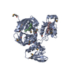
| ||||||||
|---|---|---|---|---|---|---|---|---|---|
| 1 | x 24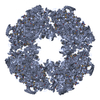
| ||||||||
| 2 | 
| ||||||||
| 3 | 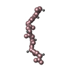
| ||||||||
| Unit cell |
|
-Components
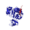
| #1: Protein | Mass: 47509.449 Da / Num. of mol.: 1 / Fragment: DegP, UNP residues 27-474 Source method: isolated from a genetically manipulated source Source: (gene. exp.) References: UniProt: P0C0V0, Hydrolases; Acting on peptide bonds (peptidases); Serine endopeptidases |
|---|---|
| #2: Protein/peptide | Mass: 443.539 Da / Num. of mol.: 1 / Source method: obtained synthetically / Production host: |
| #3: Protein/peptide | Mass: 613.749 Da / Num. of mol.: 1 / Source method: obtained synthetically |
| Has protein modification | Y |
-Experimental details
-Experiment
| Experiment | Method: X-RAY DIFFRACTION / Number of used crystals: 1 |
|---|
- Sample preparation
| Crystal | Density Matthews: 3.51 Å3/Da / Density % sol: 64.97 % |
|---|
-Data collection
| Radiation | Scattering type: x-ray |
|---|---|
| Radiation wavelength | Relative weight: 1 |
- Processing
| Software |
| ||||||||||||||||||||||||||||||||||||||||||
|---|---|---|---|---|---|---|---|---|---|---|---|---|---|---|---|---|---|---|---|---|---|---|---|---|---|---|---|---|---|---|---|---|---|---|---|---|---|---|---|---|---|---|---|
| Refinement | Method to determine structure: SAD / Resolution: 3→14.963 Å / Occupancy max: 1 / Occupancy min: 1 / FOM work R set: 0.8461 / SU ML: 0.33 / σ(F): 0 / Stereochemistry target values: Engh & Huber
| ||||||||||||||||||||||||||||||||||||||||||
| Solvent computation | Shrinkage radii: 0.9 Å / VDW probe radii: 1.11 Å / Solvent model: FLAT BULK SOLVENT MODEL / Bsol: 39.181 Å2 / ksol: 0.302 e/Å3 | ||||||||||||||||||||||||||||||||||||||||||
| Displacement parameters | Biso max: 218.7 Å2 / Biso mean: 74.1252 Å2 / Biso min: 13.04 Å2
| ||||||||||||||||||||||||||||||||||||||||||
| Refinement step | Cycle: LAST / Resolution: 3→14.963 Å
| ||||||||||||||||||||||||||||||||||||||||||
| Refine LS restraints |
| ||||||||||||||||||||||||||||||||||||||||||
| LS refinement shell | Refine-ID: X-RAY DIFFRACTION / Total num. of bins used: 5
|
