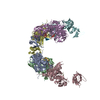
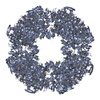
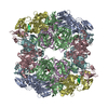
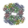
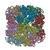
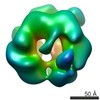
[English] 日本語
Yorodumi
- PDB-3otp: Crystal structure of the DegP dodecamer with a model substrate -

+
Open data


ID or keywords:

Loading...

-
Basic information

Entry
Database: PDB / ID: 3otp
TitleCrystal structure of the DegP dodecamer with a model substrate
Components
  • Lysozyme C
  • Protease do
KeywordsHYDROLASE / typsin-like protease domain PDZ domains / protease
Function / homology
Function and homology information


peptidase Do / programmed cell death / response to temperature stimulus / protein quality control for misfolded or incompletely synthesized proteins / chaperone-mediated protein folding / serine-type peptidase activity / Lactose synthesis / Antimicrobial peptides / Neutrophil degranulation / beta-N-acetylglucosaminidase activity ...peptidase Do / programmed cell death / response to temperature stimulus / protein quality control for misfolded or incompletely synthesized proteins / chaperone-mediated protein folding / serine-type peptidase activity / Lactose synthesis / Antimicrobial peptides / Neutrophil degranulation / beta-N-acetylglucosaminidase activity / cell wall macromolecule catabolic process / protein folding / lysozyme / lysozyme activity / peptidase activity / response to heat / outer membrane-bounded periplasmic space / defense response to Gram-negative bacterium / killing of cells of another organism / response to oxidative stress / periplasmic space / defense response to Gram-positive bacterium / defense response to bacterium / positive regulation of apoptotic process / serine-type endopeptidase activity / endoplasmic reticulum / proteolysis / extracellular space / identical protein binding / plasma membrane / cytoplasm
Similarity search - Function
Peptidase S1C, Do / Peptidase S1C / Trypsin-like peptidase domain / Thrombin, subunit H - #120 / PDZ domain / Pdz3 Domain / PDZ domain / PDZ domain profile. / Glycoside hydrolase, family 22, lysozyme / Domain present in PSD-95, Dlg, and ZO-1/2. ...Peptidase S1C, Do / Peptidase S1C / Trypsin-like peptidase domain / Thrombin, subunit H - #120 / PDZ domain / Pdz3 Domain / PDZ domain / PDZ domain profile. / Glycoside hydrolase, family 22, lysozyme / Domain present in PSD-95, Dlg, and ZO-1/2. / PDZ domain / Glycoside hydrolase family 22 domain / Glycosyl hydrolases family 22 (GH22) domain signature. / Glycoside hydrolase, family 22 / C-type lysozyme/alpha-lactalbumin family / Glycosyl hydrolases family 22 (GH22) domain profile. / Alpha-lactalbumin / lysozyme C / PDZ superfamily / Lysozyme-like domain superfamily / Trypsin-like serine proteases / Thrombin, subunit H / Roll / Peptidase S1, PA clan / Beta Barrel / Mainly Beta
Similarity search - Domain/homology
Lysozyme C / Lysozyme C / Periplasmic serine endoprotease DegP
Similarity search - Component
Biological speciesEscherichia coli (E. coli)
Gallus gallus (chicken)
MethodX-RAY DIFFRACTION / SYNCHROTRON / MOLECULAR REPLACEMENT / Resolution: 3.76 Å
AuthorsKim, S. / Grant, R.A. / Sauer, R.T.
CitationJournal: Cell(Cambridge,Mass.) / Year: 2011
Title: Covalent Linkage of Distinct Substrate Degrons Controls Assembly and Disassembly of DegP Proteolytic Cages.
Authors: Kim, S. / Grant, R.A. / Sauer, R.T.
History
DepositionSep 13, 2010Deposition site: RCSB / Processing site: RCSB
Revision 1.0Jan 19, 2011Provider: repository / Type: Initial release
Revision 1.1Jul 13, 2011Group: Version format compliance
Revision 1.2Nov 8, 2017Group: Refinement description / Category: software
Item: _software.classification / _software.contact_author ..._software.classification / _software.contact_author / _software.contact_author_email / _software.date / _software.language / _software.location / _software.name / _software.type / _software.version
Revision 1.3Feb 21, 2024Group: Data collection / Database references
Category: chem_comp_atom / chem_comp_bond ...chem_comp_atom / chem_comp_bond / database_2 / struct_ref_seq_dif
Item: _database_2.pdbx_DOI / _database_2.pdbx_database_accession / _struct_ref_seq_dif.details

-
Structure visualization

Structure viewerMolecule:
MolmilJmol/JSmol

Downloads & links

-
Assembly

Deposited unit
A: Protease do
B: Protease do
C: Protease do
D: Protease do
E: Protease do
F: Protease do
G: Lysozyme C
H: Lysozyme C
I: Lysozyme C
J: Lysozyme C
K: Lysozyme C
L: Lysozyme C


Theoretical massNumber of molelcules
Total (without water)317,67212
Polymers317,67212
Non-polymers00
Water00
1
A: Protease do
B: Protease do
C: Protease do
D: Protease do
E: Protease do
F: Protease do
G: Lysozyme C
H: Lysozyme C
I: Lysozyme C
J: Lysozyme C
K: Lysozyme C
L: Lysozyme C

A: Protease do
B: Protease do
C: Protease do
D: Protease do
E: Protease do
F: Protease do
G: Lysozyme C
H: Lysozyme C
I: Lysozyme C
J: Lysozyme C
K: Lysozyme C
L: Lysozyme C


Theoretical massNumber of molelcules
Total (without water)635,34424
Polymers635,34424
Non-polymers00
Water0
TypeNameSymmetry operationNumber
identity operation1_555x,y,z1
crystal symmetry operation2_556-x,y,-z+11
Buried area59540 Å2
ΔGint-396 kcal/mol
Surface area189770 Å2
Unit cell
Length a, b, c (Å)215.427, 122.758, 138.739
Angle α, β, γ (deg.)90.000, 117.980, 90.000
Int Tables number5
Space group name H-MC121
Noncrystallographic symmetry (NCS)NCS domain:
IDEns-IDDetails
11A
21B
31C
41D
51E
61F
12A
22B
32C
42D
52E
62F
13B
23D
33E
14A
24F
34C
15G
25H
35I
45J
55K
65L
16G
26H
36I
46J
56K
66L

NCS domain segments:
Dom-IDComponent-IDEns-IDSelection detailsAuth asym-IDAuth seq-ID
111chain A and resseq 11:263A11 - 263
211chain B and resseq 11:263B11 - 263
311chain C and resseq 11:263C11 - 263
411chain D and resseq 11:263D11 - 263
511chain E and resseq 11:263E11 - 263
611chain F and resseq 11:263F11 - 263
112chain A and resseq 264:352A264 - 352
212chain B and resseq 264:352B264 - 352
312chain C and resseq 264:352C264 - 352
412chain D and resseq 264:352D264 - 352
512chain E and resseq 264:352E264 - 352
612chain F and resseq 264:352F264 - 352
113chain B and resseq 363:448B363 - 448
213chain D and resseq 363:448D363 - 448
313chain E and resseq 363:448E363 - 448
114chain A and resseq 368:448A368 - 448
214chain F and resseq 368:448F368 - 448
314chain C and resseq 368:448C368 - 448
115chain G and resseq 54:58G54 - 58
215chain H and resseq 54:58H54 - 58
315chain I and resseq 54:58I54 - 58
415chain J and resseq 54:58J54 - 58
515chain K and resseq 54:58K54 - 58
615chain L and resseq 54:58L54 - 58
116chain G and resseq 28:33G28 - 33
216chain H and resseq 28:33H28 - 33
316chain I and resseq 28:33I28 - 33
416chain J and resseq 28:33J28 - 33
516chain K and resseq 28:33K28 - 33
616chain L and resseq 28:33L28 - 33

NCS ensembles :
ID
1
2
3
4
5
6

-
Components

#1: Protein
Protease do


Mass: 48137.301 Da / Num. of mol.: 6 / Fragment: UNP residues 27-474 / Mutation: S210A
Source method: isolated from a genetically manipulated source
Source: (gene. exp.) Escherichia coli (E. coli) / Gene: b0161, degP, htrA, JW0157, ptd / Production host: Escherichia coli (E. coli)
References: UniProt: P0C0V0, Hydrolases; Acting on peptide bonds (peptidases); Serine endopeptidases
#2: Protein/peptide
Lysozyme C


Mass: 4808.048 Da / Num. of mol.: 6 / Fragment: a model peptide substrate / Mutation: C30S
Source method: isolated from a genetically manipulated source
Source: (gene. exp.) Gallus gallus (chicken) / Gene: lysozyme C, LYZ / Production host: Escherichia coli (E. coli) / References: UniProt: B8YK79, UniProt: P00698*PLUS, lysozyme

-
Experimental details

-
Experiment

ExperimentMethod: X-RAY DIFFRACTION / Number of used crystals: 1

-
Sample preparation

CrystalDensity Matthews: 2.55 Å3/Da / Density % sol: 51.76 %
Crystal growTemperature: 291 K / Method: vapor diffusion, hanging drop / pH: 3.6
Details: 65 mM citric acid, 35 mM Bis-Tris propane, 8% PEG3350, pH 3.6, VAPOR DIFFUSION, HANGING DROP, temperature 18K, temperature 291K

-
Data collection

DiffractionMean temperature: 100 K
Diffraction sourceSource: SYNCHROTRON / Site: APS / Beamline: 24-ID-C / Wavelength: 0.97918 Å
DetectorType: ADSC QUANTUM 315 / Detector: CCD / Date: Apr 19, 2010 / Details: crystal monochromator
RadiationMonochromator: crystal monochromator / Protocol: SINGLE WAVELENGTH / Monochromatic (M) / Laue (L): M / Scattering type: x-ray
Radiation wavelengthWavelength: 0.97918 Å / Relative weight: 1
ReflectionResolution: 3.76→100 Å / Num. all: 28836 / Num. obs: 28836 / % possible obs: 90.8 % / Observed criterion σ(F): 0 / Observed criterion σ(I): 0 / Redundancy: 2.9 % / Rsym value: 0.125 / Χ2: 1.117 / Net I/σ(I): 5.3
Reflection shell
Resolution (Å)Redundancy (%)Rmerge(I) obsNum. unique allΧ2% possible all
3.76-3.942.90.68828420.73890.5
3.94-4.092.90.52929010.79291.7
4.09-4.282.90.33428840.84291.1
4.28-4.512.90.22528641.01291.3
4.51-4.7930.18629071.05491.6
4.79-5.1630.1728981.04991.7
5.16-5.6830.1728771.190.9
5.68-6.530.13828961.19591.1
6.5-8.192.90.08728881.40790.4
8.19-10030.04728791.96588.5

-
Processing

Software
NameVersionClassificationNB
DENZOdata reduction
SCALEPACKdata scaling
PHENIX1.6.4_486refinement
PDB_EXTRACT3.1data extraction
ADSCQuantumdata collection
HKL-2000data reduction
PHASERphasing
RefinementMethod to determine structure: MOLECULAR REPLACEMENT / Resolution: 3.76→14.991 Å / Occupancy max: 1 / Occupancy min: 1 / FOM work R set: 0.8412 / SU ML: 0.37 / σ(F): 1.34 / Stereochemistry target values: ML
RfactorNum. reflection% reflectionSelection details
Rfree0.2506 1369 4.83 %random
Rwork0.2187 ---
all0.2202 28349 --
obs0.2202 28349 88.25 %-
Solvent computationShrinkage radii: 0.9 Å / VDW probe radii: 1.11 Å / Solvent model: FLAT BULK SOLVENT MODEL / Bsol: 55.003 Å2 / ksol: 0.268 e/Å3
Displacement parametersBiso max: 241.85 Å2 / Biso mean: 123.4489 Å2 / Biso min: 37.43 Å2
Baniso -1Baniso -2Baniso -3
1-0.0874 Å2-0 Å2-0.5255 Å2
2--20.3638 Å20 Å2
3----20.4512 Å2
Refinement stepCycle: LAST / Resolution: 3.76→14.991 Å
ProteinNucleic acidLigandSolventTotal
Num. atoms17037 0 0 0 17037
Refine LS restraints
Refine-IDTypeDev idealNumber
X-RAY DIFFRACTIONf_bond_d0.00317173
X-RAY DIFFRACTIONf_angle_d0.61323226
X-RAY DIFFRACTIONf_chiral_restr0.0382836
X-RAY DIFFRACTIONf_plane_restr0.0033065
X-RAY DIFFRACTIONf_dihedral_angle_d10.4016251
Refine LS restraints NCS
Ens-IDDom-IDAuth asym-IDNumberRefine-IDTypeRms dev position (Å)
11A1483X-RAY DIFFRACTIONPOSITIONAL0.218
12B1483X-RAY DIFFRACTIONPOSITIONAL0.218
13C1477X-RAY DIFFRACTIONPOSITIONAL0.229
14D1472X-RAY DIFFRACTIONPOSITIONAL0.351
15E1479X-RAY DIFFRACTIONPOSITIONAL0.115
16F1479X-RAY DIFFRACTIONPOSITIONAL0.145
21A625X-RAY DIFFRACTIONPOSITIONAL0.222
22B625X-RAY DIFFRACTIONPOSITIONAL0.222
23C629X-RAY DIFFRACTIONPOSITIONAL0.162
24D621X-RAY DIFFRACTIONPOSITIONAL0.202
25E621X-RAY DIFFRACTIONPOSITIONAL0.101
26F625X-RAY DIFFRACTIONPOSITIONAL0.099
31B610X-RAY DIFFRACTIONPOSITIONAL0.299
32D610X-RAY DIFFRACTIONPOSITIONAL0.299
33E602X-RAY DIFFRACTIONPOSITIONAL0.177
41A561X-RAY DIFFRACTIONPOSITIONAL0.498
42F561X-RAY DIFFRACTIONPOSITIONAL0.498
43C561X-RAY DIFFRACTIONPOSITIONAL0.234
51G38X-RAY DIFFRACTIONPOSITIONAL0.063
52H38X-RAY DIFFRACTIONPOSITIONAL0.063
53I38X-RAY DIFFRACTIONPOSITIONAL0.169
54J38X-RAY DIFFRACTIONPOSITIONAL0.073
55K38X-RAY DIFFRACTIONPOSITIONAL0.05
56L38X-RAY DIFFRACTIONPOSITIONAL0.062
61G28X-RAY DIFFRACTIONPOSITIONAL0.039
62H28X-RAY DIFFRACTIONPOSITIONAL0.039
63I28X-RAY DIFFRACTIONPOSITIONAL0.044
64J28X-RAY DIFFRACTIONPOSITIONAL0.033
65K28X-RAY DIFFRACTIONPOSITIONAL0.044
66L28X-RAY DIFFRACTIONPOSITIONAL0.041
LS refinement shell

Refine-ID: X-RAY DIFFRACTION / Total num. of bins used: 10

Resolution (Å)Rfactor RfreeNum. reflection RfreeRfactor RworkNum. reflection RworkNum. reflection all% reflection obs (%)
3.76-3.8910.293960.29661890198663
3.891-4.04390.24951410.2732762290391
4.0439-4.2240.27741520.24752781293391
4.224-4.44120.23231340.21542782291691
4.4412-4.71130.24551450.20412782292792
4.7113-5.0620.2461330.20212804293792
5.062-5.54790.22461470.20422807295491
5.5479-6.2980.2771490.23932785293491
6.298-7.7480.26481440.23112798294291
7.748-14.99110.2371280.19212789291789

+
About Yorodumi

-
News

-
Feb 9, 2022. New format data for meta-information of EMDB entries

New format data for meta-information of EMDB entries

  • Version 3 of the EMDB header file is now the official format.
  • The previous official version 1.9 will be removed from the archive.

Related info.:EMDB header

External links:wwPDB to switch to version 3 of the EMDB data model

-
Aug 12, 2020. Covid-19 info

Covid-19 info

URL: https://pdbjlvh1.pdbj.org/emnavi/covid19.php

New page: Covid-19 featured information page in EM Navigator.

Related info.:Covid-19 info / Mar 5, 2020. Novel coronavirus structure data

+
Mar 5, 2020. Novel coronavirus structure data

Novel coronavirus structure data

Related info.:Yorodumi Speices / Aug 12, 2020. Covid-19 info

External links:COVID-19 featured content - PDBj / Molecule of the Month (242):Coronavirus Proteases

+
Jan 31, 2019. EMDB accession codes are about to change! (news from PDBe EMDB page)

EMDB accession codes are about to change! (news from PDBe EMDB page)

  • The allocation of 4 digits for EMDB accession codes will soon come to an end. Whilst these codes will remain in use, new EMDB accession codes will include an additional digit and will expand incrementally as the available range of codes is exhausted. The current 4-digit format prefixed with “EMD-” (i.e. EMD-XXXX) will advance to a 5-digit format (i.e. EMD-XXXXX), and so on. It is currently estimated that the 4-digit codes will be depleted around Spring 2019, at which point the 5-digit format will come into force.
  • The EM Navigator/Yorodumi systems omit the EMD- prefix.

Related info.:Q: What is EMD? / ID/Accession-code notation in Yorodumi/EM Navigator

External links:EMDB Accession Codes are Changing Soon! / Contact to PDBj

+
Jul 12, 2017. Major update of PDB

Major update of PDB

  • wwPDB released updated PDB data conforming to the new PDBx/mmCIF dictionary.
  • This is a major update changing the version number from 4 to 5, and with Remediation, in which all the entries are updated.
  • In this update, many items about electron microscopy experimental information are reorganized (e.g. em_software).
  • Now, EM Navigator and Yorodumi are based on the updated data.

External links:wwPDB Remediation / Enriched Model Files Conforming to OneDep Data Standards Now Available in the PDB FTP Archive

-
Yorodumi

Thousand views of thousand structures

  • Yorodumi is a browser for structure data from EMDB, PDB, SASBDB, etc.
  • This page is also the successor to EM Navigator detail page, and also detail information page/front-end page for Omokage search.
  • The word "yorodu" (or yorozu) is an old Japanese word meaning "ten thousand". "mi" (miru) is to see.

Related info.:EMDB / PDB / SASBDB / Comparison of 3 databanks / Yorodumi Search / Aug 31, 2016. New EM Navigator & Yorodumi / Yorodumi Papers / Jmol/JSmol / Function and homology information / Changes in new EM Navigator and Yorodumi

Read more