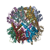
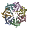
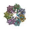
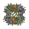
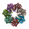
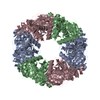
DNA BINDING PROTEIN / Helicase / recombination / DNA-binding protein / Lon-protease
Function / homology
Function and homology information
recombinational repair / ATP-dependent DNA damage sensor activity / Hydrolases; Acting on acid anhydrides; Acting on acid anhydrides to facilitate cellular and subcellular movement / damaged DNA binding / hydrolase activity / ATP binding / metal ion binding Similarity search - Function
DNA repair protein RadA / Subunit ChlI of Mg-chelatase / AAA domain / DNA recombination and repair protein RecA-like, ATP-binding domain / RecA family profile 1. / Ribosomal protein S5 domain 2-type fold, subgroup / Ribosomal protein S5 domain 2-type fold / ATPases associated with a variety of cellular activities / AAA+ ATPase domain / P-loop containing nucleoside triphosphate hydrolase Similarity search - Domain/homology
Resolution: 3.5→49.21 Å / Cor.coef. Fo:Fc: 0.949 / Cor.coef. Fo:Fc free: 0.945 / SU B: 51.177 / SU ML: 0.366 / Cross valid method: THROUGHOUT / ESU R Free: 0.531 / Details: HYDROGENS HAVE BEEN ADDED IN THE RIDING POSITIONS
Rfactor
Num. reflection
% reflection
Selection details
Rfree
0.22398
1022
5 %
RANDOM
Rwork
0.1829
-
-
-
obs
0.18497
19401
99.94 %
-
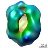
Solvent computation
Ion probe radii: 1 Å / Shrinkage radii: 1 Å / VDW probe radii: 1.1 Å
 Movie
Movie Controller


 Open data
 Basic information
 Components
 Keywords
 Function and homology information
 Streptococcus pneumoniae (bacteria)
 X-RAY DIFFRACTION /
  Authors
 France, 1items
  Citation
 Structure visualization
 Downloads & links
 Other downloads


 PDBj
PDBj
 Assembly

 Mass: 402.188 Da / Num. of mol.: 3 / Source method: obtained synthetically / Formula: C10H16N2O11P2
 Mass: 24.305 Da / Num. of mol.: 3 / Source method: obtained synthetically / Formula: Mg
 Sample preparation
 Processing