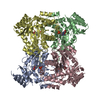
BH3-only proteins associate with and inactivate anti-apoptotic BCL-2 members / RAS processing / BH1 domain binding / regulation of development, heterochronic / positive regulation of apoptotic process involved in development / caspase complex / positive regulation of synapse pruning / peptidase activator activity involved in apoptotic process / positive regulation of protein processing / positive regulation of mitochondrial fusion ...BH3-only proteins associate with and inactivate anti-apoptotic BCL-2 members / RAS processing / BH1 domain binding / regulation of development, heterochronic / positive regulation of apoptotic process involved in development / caspase complex / positive regulation of synapse pruning / peptidase activator activity involved in apoptotic process / positive regulation of protein processing / positive regulation of mitochondrial fusion / caspase binding / embryonic morphogenesis / apoptotic process involved in development / cysteine-type endopeptidase activator activity / embryo development ending in birth or egg hatching / actin filament depolymerization / negative regulation of execution phase of apoptosis / muscle cell cellular homeostasis / regulation of cell size / negative regulation of programmed cell death / cysteine-type endopeptidase activator activity involved in apoptotic process / organelle membrane / regulation of synapse organization / BH3 domain binding / regulation of cell adhesion / endopeptidase activator activity / negative regulation of protein-containing complex assembly / mitophagy / extrinsic apoptotic signaling pathway in absence of ligand / endomembrane system / release of cytochrome c from mitochondria / GTPase activator activity / protein sequestering activity / regulation of protein stability / protein processing / ADP binding / intrinsic apoptotic signaling pathway in response to DNA damage / presynapse / channel activity / defense response to Gram-negative bacterium / perikaryon / mitochondrial outer membrane / positive regulation of apoptotic process / neuronal cell body / apoptotic process / negative regulation of apoptotic process / perinuclear region of cytoplasm / magnesium ion binding / protein-containing complex / mitochondrion / ATP binding / membrane / identical protein binding / nucleus / cytosol / cytoplasm Similarity search - Function
In the structure databanks used in Yorodumi, some data are registered as the other names, "COVID-19 virus" and "2019-nCoV". Here are the details of the virus and the list of structure data.
Jan 31, 2019. EMDB accession codes are about to change! (news from PDBe EMDB page)
EMDB accession codes are about to change! (news from PDBe EMDB page)
The allocation of 4 digits for EMDB accession codes will soon come to an end. Whilst these codes will remain in use, new EMDB accession codes will include an additional digit and will expand incrementally as the available range of codes is exhausted. The current 4-digit format prefixed with “EMD-” (i.e. EMD-XXXX) will advance to a 5-digit format (i.e. EMD-XXXXX), and so on. It is currently estimated that the 4-digit codes will be depleted around Spring 2019, at which point the 5-digit format will come into force.
The EM Navigator/Yorodumi systems omit the EMD- prefix.
Related info.:Q: What is EMD? / ID/Accession-code notation in Yorodumi/EM Navigator
Yorodumi is a browser for structure data from EMDB, PDB, SASBDB, etc.
This page is also the successor to EM Navigator detail page, and also detail information page/front-end page for Omokage search.
The word "yorodu" (or yorozu) is an old Japanese word meaning "ten thousand". "mi" (miru) is to see.
Related info.:EMDB / PDB / SASBDB / Comparison of 3 databanks / Yorodumi Search / Aug 31, 2016. New EM Navigator & Yorodumi / Yorodumi Papers / Jmol/JSmol / Function and homology information / Changes in new EM Navigator and Yorodumi
 Movie
Movie Controller


 Open data
 Basic information
 Components
 Keywords
 Function and homology information
 X-RAY DIFFRACTION /
  Authors
 Citation
 Structure visualization
 Downloads & links
 Other downloads
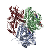

 PDBj
PDBj


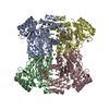
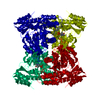
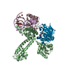
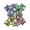
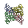
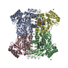
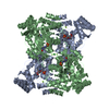
 Assembly
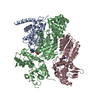


 Mass: 24.305 Da / Num. of mol.: 2 / Source method: obtained synthetically / Formula: Mg
 Mass: 507.181 Da / Num. of mol.: 2 / Source method: obtained synthetically / Formula: C10H16N5O13P3 / Comment: ATP, energy-carrying molecule*YM
 Mass: 18.015 Da / Num. of mol.: 260 / Source method: isolated from a natural source / Formula: H2O
 Sample preparation
 Processing