 Movie
Movie Controller


+ Open data

- Basic information
| Entry | Database: PDB / ID: 6jw6 | ||||||
|---|---|---|---|---|---|---|---|
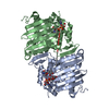
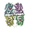
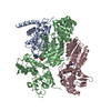
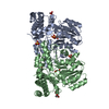
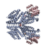
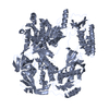
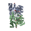
| Title | The crystal structure of KanD2 in complex with NAD | ||||||
 Components Components | Dehydrogenase | ||||||
 Keywords Keywords | OXIDOREDUCTASE / Rossmann fold / NAD binding | ||||||
| Function / homology |  Function and homology information Function and homology information | ||||||
| Biological species |  Streptomyces kanamyceticus (bacteria) Streptomyces kanamyceticus (bacteria) | ||||||
| Method |  X-RAY DIFFRACTION / SYNCHROTRON / MOLECULAR REPLACEMENT / Resolution: 2.8 Å X-RAY DIFFRACTION / SYNCHROTRON / MOLECULAR REPLACEMENT / Resolution: 2.8 Å | ||||||
 Authors Authors | Kudo, F. / Kitayama, Y. / Miyanaga, A. / Hirayama, A. / Eguchi, T. | ||||||
 Citation Citation | Journal: Biochemistry / Year: 2020 Title: Biochemical and structural analysis of a dehydrogenase, KanD2, and an aminotransferase, KanS2, that are responsible for the construction of the kanosamine moiety in kanamycin biosynthesis. Authors: Kudo, F. / Kitayama, Y. / Miyanaga, A. / Hirayama, A. / Eguchi, T. | ||||||
| History |
|
- Structure visualization
| Structure viewer | Molecule: MolmilJmol/JSmol |
|---|
- Downloads & links
-Download
| PDBx/mmCIF format | 6jw6.cif.gz | 275.9 KB | Display | PDBx/mmCIF format |
|---|---|---|---|---|
| PDB format | pdb6jw6.ent.gz | 224.7 KB | Display | PDB format |
| PDBx/mmJSON format | 6jw6.json.gz | Tree view | PDBx/mmJSON format | |
| Others |  Other downloads Other downloads |
-Validation report
| Arichive directory | https://data.pdbj.org/pub/pdb/validation_reports/jw/6jw6ftp://data.pdbj.org/pub/pdb/validation_reports/jw/6jw6 | HTTPS FTP |
|---|
-Related structure data
| Related structure data |  6jw7C  6jw8C  4h3vS S: Starting model for refinement C: citing same article ( |
|---|---|
| Similar structure data |

-Links
 PDBj
PDBj- Assembly
| Deposited unit | 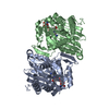
| ||||||||||||||||||
|---|---|---|---|---|---|---|---|---|---|---|---|---|---|---|---|---|---|---|---|
| 1 | 
| ||||||||||||||||||
| Unit cell |
| ||||||||||||||||||
| Noncrystallographic symmetry (NCS) | NCS domain:
NCS domain segments: Component-ID: _ / Ens-ID: 1 / Beg auth comp-ID: PRO / Beg label comp-ID: PRO / End auth comp-ID: VAL / End label comp-ID: VAL / Refine code: _ / Auth seq-ID: 4 - 361 / Label seq-ID: 4 - 361
|
-Components
| #1: Protein | Mass: 41108.059 Da / Num. of mol.: 2 Source method: isolated from a genetically manipulated source Source: (gene. exp.) Streptomyces kanamyceticus (bacteria) / Plasmid: pET30 / Production host: References: UniProt: Q6L737, Oxidoreductases; Acting on the CH-OH group of donors; With NAD+ or NADP+ as acceptor #2: Chemical |   Mass: 663.425 Da / Num. of mol.: 2 / Source method: obtained synthetically / Formula: C21H27N7O14P2 / Feature type: SUBJECT OF INVESTIGATION / Comment: NAD*YM Mass: 663.425 Da / Num. of mol.: 2 / Source method: obtained synthetically / Formula: C21H27N7O14P2 / Feature type: SUBJECT OF INVESTIGATION / Comment: NAD*YM#3: Water | ChemComp-HOH / |  Mass: 18.015 Da / Num. of mol.: 8 / Source method: isolated from a natural source / Formula: H2O Mass: 18.015 Da / Num. of mol.: 8 / Source method: isolated from a natural source / Formula: H2OHas ligand of interest | Y | |
|---|
-Experimental details
-Experiment
| Experiment | Method: X-RAY DIFFRACTION / Number of used crystals: 1 |
|---|
- Sample preparation
| Crystal | Density Matthews: 3.39 Å3/Da / Density % sol: 63.73 % |
|---|---|
| Crystal grow | Temperature: 293 K / Method: vapor diffusion, sitting drop / pH: 8 / Details: PEG 8000, imidazole, calcium acetate, NAD |
-Data collection
| Diffraction | Mean temperature: 100 K / Serial crystal experiment: N |
|---|---|
| Diffraction source | Source: SYNCHROTRON / Site: Photon Factory  / Beamline: AR-NW12A / Wavelength: 1 Å / Beamline: AR-NW12A / Wavelength: 1 Å |
| Detector | Type: ADSC QUANTUM 270 / Detector: CCD / Date: Nov 21, 2017 |
| Radiation | Protocol: SINGLE WAVELENGTH / Monochromatic (M) / Laue (L): M / Scattering type: x-ray |
| Radiation wavelength | Wavelength: 1 Å / Relative weight: 1 |
| Reflection | Resolution: 2.8→50 Å / Num. obs: 27212 / % possible obs: 100 % / Redundancy: 11.1 % / Rmerge(I) obs: 0.085 / Net I/σ(I): 23.3 |
| Reflection shell | Resolution: 2.8→2.95 Å / Redundancy: 11.3 % / Rmerge(I) obs: 0.918 / Mean I/σ(I) obs: 2.5 / Num. unique obs: 3916 / % possible all: 100 |
- Processing
| Software |
| |||||||||||||||||||||||||||||||||||||||||||||||||||||||||||||||||||||||||||
|---|---|---|---|---|---|---|---|---|---|---|---|---|---|---|---|---|---|---|---|---|---|---|---|---|---|---|---|---|---|---|---|---|---|---|---|---|---|---|---|---|---|---|---|---|---|---|---|---|---|---|---|---|---|---|---|---|---|---|---|---|---|---|---|---|---|---|---|---|---|---|---|---|---|---|---|---|
| Refinement | Method to determine structure: MOLECULAR REPLACEMENT Starting model: 4H3V Resolution: 2.8→48.18 Å / Cor.coef. Fo:Fc: 0.955 / Cor.coef. Fo:Fc free: 0.929 / SU B: 26.569 / SU ML: 0.231 / Cross valid method: THROUGHOUT / σ(F): 0 / ESU R: 0.595 / ESU R Free: 0.315 / Stereochemistry target values: MAXIMUM LIKELIHOOD Details: U VALUES : WITH TLS ADDED HYDROGENS HAVE BEEN ADDED IN THE RIDING POSITIONS
| |||||||||||||||||||||||||||||||||||||||||||||||||||||||||||||||||||||||||||
| Solvent computation | Ion probe radii: 0.8 Å / Shrinkage radii: 0.8 Å / VDW probe radii: 1.2 Å / Solvent model: MASK | |||||||||||||||||||||||||||||||||||||||||||||||||||||||||||||||||||||||||||
| Displacement parameters | Biso max: 176.51 Å2 / Biso mean: 79.263 Å2 / Biso min: 44.62 Å2
| |||||||||||||||||||||||||||||||||||||||||||||||||||||||||||||||||||||||||||
| Refinement step | Cycle: final / Resolution: 2.8→48.18 Å
| |||||||||||||||||||||||||||||||||||||||||||||||||||||||||||||||||||||||||||
| Refine LS restraints |
| |||||||||||||||||||||||||||||||||||||||||||||||||||||||||||||||||||||||||||
| Refine LS restraints NCS | Ens-ID: 1 / Number: 20175 / Refine-ID: X-RAY DIFFRACTION / Type: interatomic distance / Rms dev position: 0.08 Å / Weight position: 0.05
| |||||||||||||||||||||||||||||||||||||||||||||||||||||||||||||||||||||||||||
| LS refinement shell | Resolution: 2.8→2.873 Å / Rfactor Rfree error: 0 / Total num. of bins used: 20
| |||||||||||||||||||||||||||||||||||||||||||||||||||||||||||||||||||||||||||
| Refinement TLS params. | Method: refined / Refine-ID: X-RAY DIFFRACTION
| |||||||||||||||||||||||||||||||||||||||||||||||||||||||||||||||||||||||||||
| Refinement TLS group |
|
