 Movie
Movie Controller

[English] 日本語
 Yorodumi
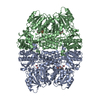
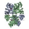
- PDB-2z9v: Crystal structure of pyridoxamine-pyruvate aminotransferase compl... -
+ Open data

- Basic information
| Entry | Database: PDB / ID: 2z9v | ||||||
|---|---|---|---|---|---|---|---|
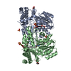
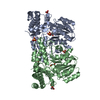
| Title | Crystal structure of pyridoxamine-pyruvate aminotransferase complexed with pyridoxamine | ||||||
 Components Components | Aspartate aminotransferase | ||||||
 Keywords Keywords | TRANSFERASE / AMINOTRANSFERASE / PYRIDOXAMINE / PYRUVATE | ||||||
| Function / homology |  Function and homology information Function and homology informationpyridoxamine-pyruvate transaminase / pyridoxamine:pyruvate transaminase activity / L-serine:pyruvate transaminase activity / : / L-alanine:glyoxylate transaminase activity / transaminase activity / pyridoxal phosphate binding Similarity search - Function | ||||||
| Biological species |  Mesorhizobium loti (bacteria) Mesorhizobium loti (bacteria) | ||||||
| Method |  X-RAY DIFFRACTION / SYNCHROTRON / MOLECULAR REPLACEMENT / Resolution: 1.7 Å X-RAY DIFFRACTION / SYNCHROTRON / MOLECULAR REPLACEMENT / Resolution: 1.7 Å | ||||||
 Authors Authors | Yoshikane, Y. / Yokochi, N. / Yamasaki, M. / Mizutani, K. / Ohnishi, K. / Mikami, B. / Hayashi, H. / Yagi, T. | ||||||
 Citation Citation | Journal: J.Biol.Chem. / Year: 2008 Title: Crystal structure of pyridoxamine-pyruvate aminotransferase from Mesorhizobium loti MAFF303099 Authors: Yoshikane, Y. / Yokochi, N. / Yamasaki, M. / Mizutani, K. / Ohnishi, K. / Mikami, B. / Hayashi, H. / Yagi, T. | ||||||
| History |
|
- Structure visualization
| Structure viewer | Molecule: MolmilJmol/JSmol |
|---|
- Downloads & links
-Download
| PDBx/mmCIF format | 2z9v.cif.gz | 172.6 KB | Display | PDBx/mmCIF format |
|---|---|---|---|---|
| PDB format | pdb2z9v.ent.gz | 137.3 KB | Display | PDB format |
| PDBx/mmJSON format | 2z9v.json.gz | Tree view | PDBx/mmJSON format | |
| Others |  Other downloads Other downloads |
-Validation report
| Arichive directory | https://data.pdbj.org/pub/pdb/validation_reports/z9/2z9vftp://data.pdbj.org/pub/pdb/validation_reports/z9/2z9v | HTTPS FTP |
|---|
-Related structure data
| Related structure data |  2z9uSC  2z9wC  2z9xC S: Starting model for refinement C: citing same article ( |
|---|---|
| Similar structure data |


-Links
 PDBj
PDBj- Assembly
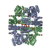
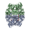
| Deposited unit | 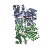
| ||||||||
|---|---|---|---|---|---|---|---|---|---|
| 1 | 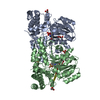
| ||||||||
| Unit cell |
|
-Components
| #1: Protein | Mass: 41503.512 Da / Num. of mol.: 2 Source method: isolated from a genetically manipulated source Source: (gene. exp.) Mesorhizobium loti (bacteria) / Strain: MAFF303099 / Gene: mlr6806 / Plasmid: PET21A / Production host: References: UniProt: Q988B8, pyridoxamine-pyruvate transaminase #2: Chemical | ChemComp-SO4 /   Mass: 96.063 Da / Num. of mol.: 5 / Source method: obtained synthetically / Formula: SO4 Mass: 96.063 Da / Num. of mol.: 5 / Source method: obtained synthetically / Formula: SO4#3: Chemical |   Mass: 168.193 Da / Num. of mol.: 2 / Source method: obtained synthetically / Formula: C8H12N2O2 Mass: 168.193 Da / Num. of mol.: 2 / Source method: obtained synthetically / Formula: C8H12N2O2#4: Chemical | ChemComp-GOL /   Mass: 92.094 Da / Num. of mol.: 4 / Source method: obtained synthetically / Formula: C3H8O3 Mass: 92.094 Da / Num. of mol.: 4 / Source method: obtained synthetically / Formula: C3H8O3#5: Water | ChemComp-HOH / |  Mass: 18.015 Da / Num. of mol.: 753 / Source method: isolated from a natural source / Formula: H2O Mass: 18.015 Da / Num. of mol.: 753 / Source method: isolated from a natural source / Formula: H2O |
|---|
-Experimental details
-Experiment
| Experiment | Method: X-RAY DIFFRACTION / Number of used crystals: 1 |
|---|
- Sample preparation
| Crystal | Density Matthews: 2.21 Å3/Da / Density % sol: 44.45 % |
|---|---|
| Crystal grow | Temperature: 277 K / Method: vapor diffusion, sitting drop / pH: 8.05 Details: 0.1M HEPES, 2M Ammonium sulfate, 10mM Pyridoxamine, pH 8.05, VAPOR DIFFUSION, SITTING DROP, temperature 277K |
-Data collection
| Diffraction | Mean temperature: 100 K |
|---|---|
| Diffraction source | Source: SYNCHROTRON / Site: SPring-8  / Beamline: BL38B1 / Wavelength: 1 Å / Beamline: BL38B1 / Wavelength: 1 Å |
| Detector | Type: RIGAKU RAXIS V / Detector: IMAGE PLATE / Date: Oct 27, 2005 |
| Radiation | Monochromator: DOUBLE-CRYSTAL MONOCHROMATOR / Protocol: SINGLE WAVELENGTH / Monochromatic (M) / Laue (L): M / Scattering type: x-ray |
| Radiation wavelength | Wavelength: 1 Å / Relative weight: 1 |
| Reflection | Resolution: 1.7→50 Å / Num. obs: 82435 / % possible obs: 98.4 % / Observed criterion σ(I): -3 / Redundancy: 5.8 % / Biso Wilson estimate: 14.99 Å2 / Rmerge(I) obs: 0.039 / Net I/σ(I): 24.4 |
| Reflection shell | Resolution: 1.7→1.76 Å / Redundancy: 3 % / Rmerge(I) obs: 0.113 / Num. unique all: 7561 / % possible all: 91.9 |
- Processing
| Software |
| ||||||||||||||||||||||||||||||||||||||||||||||||||||||||||||||||||||||
|---|---|---|---|---|---|---|---|---|---|---|---|---|---|---|---|---|---|---|---|---|---|---|---|---|---|---|---|---|---|---|---|---|---|---|---|---|---|---|---|---|---|---|---|---|---|---|---|---|---|---|---|---|---|---|---|---|---|---|---|---|---|---|---|---|---|---|---|---|---|---|---|
| Refinement | Method to determine structure: MOLECULAR REPLACEMENT Starting model: PDB ID 2Z9U Resolution: 1.7→14.94 Å / Cor.coef. Fo:Fc: 0.963 / Cor.coef. Fo:Fc free: 0.952 / SU B: 1.636 / SU ML: 0.056 / Cross valid method: THROUGHOUT / ESU R: 0.099 / ESU R Free: 0.096 / Stereochemistry target values: MAXIMUM LIKELIHOOD / Details: HYDROGENS HAVE BEEN ADDED IN THE RIDING POSITIONS
| ||||||||||||||||||||||||||||||||||||||||||||||||||||||||||||||||||||||
| Solvent computation | Ion probe radii: 0.8 Å / Shrinkage radii: 0.8 Å / VDW probe radii: 1.2 Å / Solvent model: MASK | ||||||||||||||||||||||||||||||||||||||||||||||||||||||||||||||||||||||
| Displacement parameters | Biso mean: 14.852 Å2
| ||||||||||||||||||||||||||||||||||||||||||||||||||||||||||||||||||||||
| Refinement step | Cycle: LAST / Resolution: 1.7→14.94 Å
| ||||||||||||||||||||||||||||||||||||||||||||||||||||||||||||||||||||||
| Refine LS restraints |
| ||||||||||||||||||||||||||||||||||||||||||||||||||||||||||||||||||||||
| LS refinement shell | Resolution: 1.7→1.744 Å / Total num. of bins used: 20
|
