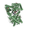
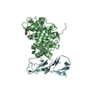
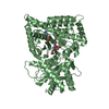
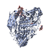
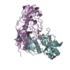
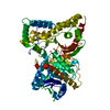
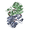
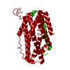
Journal: Sci Adv / Year: 2019 Title: Solving a new R2lox protein structure by microcrystal electron diffraction. Authors: Hongyi Xu / Hugo Lebrette / Max T B Clabbers / Jingjing Zhao / Julia J Griese / Xiaodong Zou / Martin Högbom / Abstract: Microcrystal electron diffraction (MicroED) has recently shown potential for structural biology. It enables the study of biomolecules from micrometer-sized 3D crystals that are too small to be ...Microcrystal electron diffraction (MicroED) has recently shown potential for structural biology. It enables the study of biomolecules from micrometer-sized 3D crystals that are too small to be studied by conventional x-ray crystallography. However, to date, MicroED has only been applied to redetermine protein structures that had already been solved previously by x-ray diffraction. Here, we present the first new protein structure-an R2lox enzyme-solved using MicroED. The structure was phased by molecular replacement using a search model of 35% sequence identity. The resulting electrostatic scattering potential map at 3.0-Å resolution was of sufficient quality to allow accurate model building and refinement. The dinuclear metal cofactor could be located in the map and was modeled as a heterodinuclear Mn/Fe center based on previous studies. Our results demonstrate that MicroED has the potential to become a widely applicable tool for revealing novel insights into protein structure and function.
Conc.: 8 mg/ml / Embedding applied: NO / Shadowing applied: NO / Staining applied: NO / Vitrification applied: YES Details: Plate-like micro-crystals grow within 48h at 21C by hanging drop vapour diffusion
In the structure databanks used in Yorodumi, some data are registered as the other names, "COVID-19 virus" and "2019-nCoV". Here are the details of the virus and the list of structure data.
Jan 31, 2019. EMDB accession codes are about to change! (news from PDBe EMDB page)
EMDB accession codes are about to change! (news from PDBe EMDB page)
The allocation of 4 digits for EMDB accession codes will soon come to an end. Whilst these codes will remain in use, new EMDB accession codes will include an additional digit and will expand incrementally as the available range of codes is exhausted. The current 4-digit format prefixed with “EMD-” (i.e. EMD-XXXX) will advance to a 5-digit format (i.e. EMD-XXXXX), and so on. It is currently estimated that the 4-digit codes will be depleted around Spring 2019, at which point the 5-digit format will come into force.
The EM Navigator/Yorodumi systems omit the EMD- prefix.
Related info.:Q: What is EMD? / ID/Accession-code notation in Yorodumi/EM Navigator
Yorodumi is a browser for structure data from EMDB, PDB, SASBDB, etc.
This page is also the successor to EM Navigator detail page, and also detail information page/front-end page for Omokage search.
The word "yorodu" (or yorozu) is an old Japanese word meaning "ten thousand". "mi" (miru) is to see.
Related info.:EMDB / PDB / SASBDB / Comparison of 3 databanks / Yorodumi Search / Aug 31, 2016. New EM Navigator & Yorodumi / Yorodumi Papers / Jmol/JSmol / Function and homology information / Changes in new EM Navigator and Yorodumi
 Movie
Movie Controller

 Yorodumi
 Open data
 Basic information
 Components
 Keywords
 Function and homology information
 Sulfolobus acidocaldarius DSM 639 (acidophilic)
 Authors
 Sweden, 4items
  Citation
 Structure visualization
 Downloads & links
 Other downloads
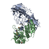


 PDBj
PDBj

 Assembly
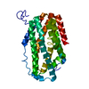
 gel filtration

 Mass: 54.938 Da / Num. of mol.: 1 / Source method: obtained synthetically / Formula: Mn
 Mass: 55.845 Da / Num. of mol.: 1 / Source method: obtained synthetically / Formula: Fe
 Sample preparation
 Processing
