 Movie
Movie Controller

[English] 日本語
 Yorodumi
- PDB-6qpi: Cryo-EM structure of calcium-free mTMEM16F lipid scramblase in na... -
+ Open data

- Basic information
| Entry | Database: PDB / ID: 6qpi | |||||||||
|---|---|---|---|---|---|---|---|---|---|---|
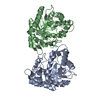
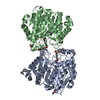
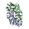
| Title | Cryo-EM structure of calcium-free mTMEM16F lipid scramblase in nanodisc | |||||||||
 Components Components | Anoctamin-6 | |||||||||
 Keywords Keywords | MEMBRANE PROTEIN / lipid scrambles / TMEM16 | |||||||||
| Function / homology |  Function and homology information Function and homology informationcalcium activated phospholipid scrambling / calcium activated galactosylceramide scrambling / calcium activated phosphatidylserine scrambling / calcium activated phosphatidylcholine scrambling / positive regulation of potassium ion export across plasma membrane / positive regulation of monoatomic ion transmembrane transport / purinergic nucleotide receptor signaling pathway / phospholipid scramblase activity / cholinergic synapse / bone mineralization involved in bone maturation ...calcium activated phospholipid scrambling / calcium activated galactosylceramide scrambling / calcium activated phosphatidylserine scrambling / calcium activated phosphatidylcholine scrambling / positive regulation of potassium ion export across plasma membrane / positive regulation of monoatomic ion transmembrane transport / purinergic nucleotide receptor signaling pathway / phospholipid scramblase activity / cholinergic synapse / bone mineralization involved in bone maturation / intracellularly calcium-gated chloride channel activity / plasma membrane phospholipid scrambling / negative regulation of cell volume / voltage-gated monoatomic ion channel activity / positive regulation of phagocytosis, engulfment / bleb assembly / Stimuli-sensing channels / calcium-activated cation channel activity / positive regulation of monocyte chemotaxis / chloride transport / dendritic cell chemotaxis / phospholipid translocation / regulation of postsynaptic membrane potential / positive regulation of bone mineralization / chloride channel complex / Neutrophil degranulation / chloride transmembrane transport / synaptic membrane / establishment of localization in cell / calcium ion transmembrane transport / blood coagulation / positive regulation of apoptotic process / protein homodimerization activity / metal ion binding / identical protein binding / plasma membrane Similarity search - Function | |||||||||
| Biological species |  | |||||||||
| Method | ELECTRON MICROSCOPY / single particle reconstruction / cryo EM / Resolution: 3.3 Å | |||||||||
 Authors Authors | Alvadia, C. / Lim, N.K. / Clerico Mosina, V. / Oostergetel, G.T. / Dutzler, R. / Paulino, C. | |||||||||
| Funding support |  Switzerland, Switzerland,  Netherlands, 2items Netherlands, 2items
| |||||||||
 Citation Citation | Journal: Elife / Year: 2019 Title: Cryo-EM structures and functional characterization of the murine lipid scramblase TMEM16F. Authors: Carolina Alvadia / Novandy K Lim / Vanessa Clerico Mosina / Gert T Oostergetel / Raimund Dutzler / Cristina Paulino / Abstract: The lipid scramblase TMEM16F initiates blood coagulation by catalyzing the exposure of phosphatidylserine in platelets. The protein is part of a family of membrane proteins, which encompasses calcium- ...The lipid scramblase TMEM16F initiates blood coagulation by catalyzing the exposure of phosphatidylserine in platelets. The protein is part of a family of membrane proteins, which encompasses calcium-activated channels for ions and lipids. Here, we reveal features of murine TMEM16F (mTMEM16F) that underlie its function as a lipid scramblase and an ion channel. The cryo-EM data of mTMEM16F in absence and presence of Ca define the ligand-free closed conformation of the protein and the structure of a Ca-bound intermediate. Both conformations resemble their counterparts of the scrambling-incompetent anion channel mTMEM16A, yet with distinct differences in the region of ion and lipid permeation. In conjunction with functional data, we demonstrate the relationship between ion conduction and lipid scrambling. Although activated by a common mechanism, both functions appear to be mediated by alternate protein conformations that are at equilibrium in the ligand-bound state. | |||||||||
| History |
|
- Structure visualization
| Movie |
Movie viewer |
|---|---|
| Structure viewer | Molecule: MolmilJmol/JSmol |
- Downloads & links
-Download
| PDBx/mmCIF format | 6qpi.cif.gz | 99.3 KB | Display | PDBx/mmCIF format |
|---|---|---|---|---|
| PDB format | pdb6qpi.ent.gz | 53.5 KB | Display | PDB format |
| PDBx/mmJSON format | 6qpi.json.gz | Tree view | PDBx/mmJSON format | |
| Others |  Other downloads Other downloads |
-Validation report
| Summary document | 6qpi_validation.pdf.gz | 975.9 KB | Display | wwPDB validaton report |
|---|---|---|---|---|
| Full document | 6qpi_full_validation.pdf.gz | 975.5 KB | Display | |
| Data in XML | 6qpi_validation.xml.gz | 23.6 KB | Display | |
| Data in CIF | 6qpi_validation.cif.gz | 35.2 KB | Display | |
| Arichive directory | https://data.pdbj.org/pub/pdb/validation_reports/qp/6qpiftp://data.pdbj.org/pub/pdb/validation_reports/qp/6qpi | HTTPS FTP |
-Related structure data
| Related structure data |  4614MC  4611C  4612C  4613C 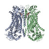 6qp6C  6qpbC  6qpcC M: map data used to model this data C: citing same article ( |
|---|---|
| Similar structure data |


-Links
 PDBj
PDBj


- Assembly
| Deposited unit | 
|
|---|---|
| 1 |
|
-Components
| #1: Protein | Mass: 106367.727 Da / Num. of mol.: 2 Source method: isolated from a genetically manipulated source Source: (gene. exp.)  Homo sapiens (human) / References: UniProt: Q6P9J9 Homo sapiens (human) / References: UniProt: Q6P9J9 |
|---|
-Experimental details
-Experiment
| Experiment | Method: ELECTRON MICROSCOPY |
|---|---|
| EM experiment | Aggregation state: PARTICLE / 3D reconstruction method: single particle reconstruction |
- Sample preparation
| Component | Name: mTMEM16F / Type: COMPLEX / Entity ID: all / Source: RECOMBINANT |
|---|---|
| Molecular weight | Value: 0.212 MDa / Experimental value: NO |
| Source (natural) | Organism: |
| Source (recombinant) | Organism: Homo sapiens (human) / Cell: HEK293T cells |
| Buffer solution | pH: 7.5 / Details: 150 mM NaCl, 20 mM HEPES, pH 7.5 and 2 mM EGTA |
| Specimen | Conc.: 1 mg/ml / Embedding applied: NO / Shadowing applied: NO / Staining applied: NO / Vitrification applied: YES |
| Specimen support | Details: at 5 mA / Grid material: GOLD / Grid mesh size: 300 divisions/in. / Grid type: Quantifoil R1.2/1.3 |
| Vitrification | Instrument: FEI VITROBOT MARK IV / Cryogen name: ETHANE / Humidity: 100 % / Chamber temperature: 288 K |
- Electron microscopy imaging
| Experimental equipment |  Model: Talos Arctica / Image courtesy: FEI Company |
|---|---|
| Microscopy | Model: FEI TALOS ARCTICA |
| Electron gun | Electron source:  FIELD EMISSION GUN / Accelerating voltage: 200 kV / Illumination mode: FLOOD BEAM FIELD EMISSION GUN / Accelerating voltage: 200 kV / Illumination mode: FLOOD BEAM |
| Electron lens | Mode: BRIGHT FIELD / Nominal magnification: 49407 X / Calibrated magnification: 49407 X / Nominal defocus max: 3000 nm / Nominal defocus min: 300 nm / Calibrated defocus min: 300 nm / Calibrated defocus max: 3000 nm / Cs: 2.7 mm / C2 aperture diameter: 100 µm / Alignment procedure: COMA FREE |
| Specimen holder | Cryogen: NITROGEN / Specimen holder model: FEI TITAN KRIOS AUTOGRID HOLDER / Temperature (max): 105 K / Temperature (min): 90 K |
| Image recording | Average exposure time: 9 sec. / Electron dose: 52 e/Å2 / Detector mode: COUNTING / Film or detector model: GATAN K2 SUMMIT (4k x 4k) / Num. of grids imaged: 10 / Num. of real images: 8706 |
| EM imaging optics | Energyfilter name: GIF Bioquantum / Energyfilter slit width: 20 eV |
| Image scans | Width: 3838 / Height: 3710 / Movie frames/image: 60 / Used frames/image: 1-60 |
- Processing
| Software | Name: PHENIX / Version: 1.14_3260: / Classification: refinement | ||||||||||||||||||||||||||||||||||||
|---|---|---|---|---|---|---|---|---|---|---|---|---|---|---|---|---|---|---|---|---|---|---|---|---|---|---|---|---|---|---|---|---|---|---|---|---|---|
| EM software |
| ||||||||||||||||||||||||||||||||||||
| CTF correction | Type: PHASE FLIPPING AND AMPLITUDE CORRECTION | ||||||||||||||||||||||||||||||||||||
| Particle selection | Num. of particles selected: 1593115 | ||||||||||||||||||||||||||||||||||||
| Symmetry | Point symmetry: C2 (2 fold cyclic) | ||||||||||||||||||||||||||||||||||||
| 3D reconstruction | Resolution: 3.3 Å / Resolution method: FSC 0.143 CUT-OFF / Num. of particles: 280891 / Algorithm: BACK PROJECTION / Num. of class averages: 9 / Symmetry type: POINT | ||||||||||||||||||||||||||||||||||||
| Atomic model building | Space: REAL |
