 Movie
Movie Controller

[English] 日本語
 Yorodumi
- PDB-7cju: Crystal structure of inactive form of chitosanase crystallized by... -
+ Open data

- Basic information
| Entry | Database: PDB / ID: 7cju | ||||||
|---|---|---|---|---|---|---|---|
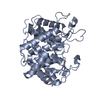
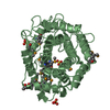
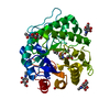
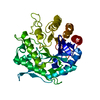
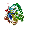
| Title | Crystal structure of inactive form of chitosanase crystallized by ammonium sulfate | ||||||
 Components Components | Glucanase | ||||||
 Keywords Keywords | HYDROLASE / enzyme | ||||||
| Function / homology |  Function and homology information Function and homology informationHydrolases; Glycosylases; Glycosidases, i.e. enzymes that hydrolyse O- and S-glycosyl compounds / polysaccharide catabolic process / hydrolase activity, hydrolyzing O-glycosyl compounds Similarity search - Function | ||||||
| Biological species |  | ||||||
| Method |  X-RAY DIFFRACTION / SYNCHROTRON / MOLECULAR REPLACEMENT / Resolution: 1.74 Å X-RAY DIFFRACTION / SYNCHROTRON / MOLECULAR REPLACEMENT / Resolution: 1.74 Å | ||||||
 Authors Authors | Guo, Y. / Qu, L. / Nishida, N. / Hoshino, T. | ||||||
| Funding support |  Japan, 1items Japan, 1items
| ||||||
 Citation Citation | Journal: Cryst.Growth Des. / Year: 2021 Title: Electrostatic Potentials around the Proteins Preferably Crystallized by Ammonium Sulfate Authors: Guo, Y. / Qu, L. / Nishida, N. / Hoshino, T. | ||||||
| History |
|
- Structure visualization
| Structure viewer | Molecule: MolmilJmol/JSmol |
|---|
- Downloads & links
-Download
| PDBx/mmCIF format | 7cju.cif.gz | 186.7 KB | Display | PDBx/mmCIF format |
|---|---|---|---|---|
| PDB format | pdb7cju.ent.gz | 144.2 KB | Display | PDB format |
| PDBx/mmJSON format | 7cju.json.gz | Tree view | PDBx/mmJSON format | |
| Others |  Other downloads Other downloads |
-Validation report
| Arichive directory | https://data.pdbj.org/pub/pdb/validation_reports/cj/7cjuftp://data.pdbj.org/pub/pdb/validation_reports/cj/7cju | HTTPS FTP |
|---|
-Related structure data
| Related structure data | 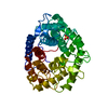 1v5cS S: Starting model for refinement |
|---|---|
| Similar structure data |


-Links
 PDBj
PDBj
- Assembly
| Deposited unit | 
| ||||||||
|---|---|---|---|---|---|---|---|---|---|
| 1 | 
| ||||||||
| 2 | 
| ||||||||
| Unit cell |
|
-Components
| #1: Protein | Mass: 44594.703 Da / Num. of mol.: 2 / Mutation: S411R,K437T Source method: isolated from a genetically manipulated source Source: (gene. exp.) References: UniProt: S3JG56, Hydrolases; Glycosylases; Glycosidases, i.e. enzymes that hydrolyse O- and S-glycosyl compounds #2: Water | ChemComp-HOH / |  Mass: 18.015 Da / Num. of mol.: 966 / Source method: isolated from a natural source / Formula: H2O Mass: 18.015 Da / Num. of mol.: 966 / Source method: isolated from a natural source / Formula: H2OSequence details | Sequence reference which is derived from Bacillus sp. K17-2 is not available at UNIPROT at the time ...Sequence reference which is derived from Bacillus sp. K17-2 is not available at UNIPROT at the time of data annotation. The sequence reference used (S3JG56) is from a different strain Bacillus cereus BAG1O-3. | |
|---|
-Experimental details
-Experiment
| Experiment | Method: X-RAY DIFFRACTION / Number of used crystals: 1 |
|---|
- Sample preparation
| Crystal | Density Matthews: 2.51 Å3/Da / Density % sol: 51.06 % / Mosaicity: 0.14 ° |
|---|---|
| Crystal grow | Temperature: 291 K / Method: vapor diffusion, sitting drop / pH: 5 / Details: 0.1M Sodium Citrate, 3.4M Ammonium Sulfate |
-Data collection
| Diffraction | Mean temperature: 100 K / Serial crystal experiment: N | ||||||||||||||||||||||||||||||
|---|---|---|---|---|---|---|---|---|---|---|---|---|---|---|---|---|---|---|---|---|---|---|---|---|---|---|---|---|---|---|---|
| Diffraction source | Source: SYNCHROTRON / Site: Photon Factory / Beamline: BL-17A / Wavelength: 1 Å | ||||||||||||||||||||||||||||||
| Detector | Type: DECTRIS EIGER X 16M / Detector: PIXEL / Date: Feb 22, 2020 / Details: mirrors | ||||||||||||||||||||||||||||||
| Radiation | Monochromator: Si(111) / Protocol: SINGLE WAVELENGTH / Monochromatic (M) / Laue (L): M / Scattering type: x-ray | ||||||||||||||||||||||||||||||
| Radiation wavelength | Wavelength: 1 Å / Relative weight: 1 | ||||||||||||||||||||||||||||||
| Reflection | Resolution: 1.739→48.441 Å / Num. obs: 90669 / % possible obs: 98.9 % / Redundancy: 7.6 % / Biso Wilson estimate: 19.9 Å2 / CC1/2: 0.996 / Rmerge(I) obs: 0.158 / Rpim(I) all: 0.061 / Rrim(I) all: 0.17 / Net I/σ(I): 8.1 | ||||||||||||||||||||||||||||||
| Reflection shell | Diffraction-ID: 1
|
- Processing
| Software |
| ||||||||||||||||||||||||||||||||||||||||||||||||||||||||||||||||||||||||||||||||||||||||||||||||||||||||||||||||||||||||||||||||||||||||||||||||||||||||||||||||||||||||||||||||||||||||||
|---|---|---|---|---|---|---|---|---|---|---|---|---|---|---|---|---|---|---|---|---|---|---|---|---|---|---|---|---|---|---|---|---|---|---|---|---|---|---|---|---|---|---|---|---|---|---|---|---|---|---|---|---|---|---|---|---|---|---|---|---|---|---|---|---|---|---|---|---|---|---|---|---|---|---|---|---|---|---|---|---|---|---|---|---|---|---|---|---|---|---|---|---|---|---|---|---|---|---|---|---|---|---|---|---|---|---|---|---|---|---|---|---|---|---|---|---|---|---|---|---|---|---|---|---|---|---|---|---|---|---|---|---|---|---|---|---|---|---|---|---|---|---|---|---|---|---|---|---|---|---|---|---|---|---|---|---|---|---|---|---|---|---|---|---|---|---|---|---|---|---|---|---|---|---|---|---|---|---|---|---|---|---|---|---|---|---|---|
| Refinement | Method to determine structure: MOLECULAR REPLACEMENT Starting model: pdbid 1V5C Resolution: 1.74→48.44 Å / SU ML: 0.19 / Cross valid method: THROUGHOUT / σ(F): 1.36 / Phase error: 17.73 / Stereochemistry target values: ML
| ||||||||||||||||||||||||||||||||||||||||||||||||||||||||||||||||||||||||||||||||||||||||||||||||||||||||||||||||||||||||||||||||||||||||||||||||||||||||||||||||||||||||||||||||||||||||||
| Solvent computation | Shrinkage radii: 0.9 Å / VDW probe radii: 1.11 Å / Solvent model: FLAT BULK SOLVENT MODEL | ||||||||||||||||||||||||||||||||||||||||||||||||||||||||||||||||||||||||||||||||||||||||||||||||||||||||||||||||||||||||||||||||||||||||||||||||||||||||||||||||||||||||||||||||||||||||||
| Displacement parameters | Biso max: 85.77 Å2 / Biso mean: 21.6695 Å2 / Biso min: 6.97 Å2 | ||||||||||||||||||||||||||||||||||||||||||||||||||||||||||||||||||||||||||||||||||||||||||||||||||||||||||||||||||||||||||||||||||||||||||||||||||||||||||||||||||||||||||||||||||||||||||
| Refinement step | Cycle: final / Resolution: 1.74→48.44 Å
| ||||||||||||||||||||||||||||||||||||||||||||||||||||||||||||||||||||||||||||||||||||||||||||||||||||||||||||||||||||||||||||||||||||||||||||||||||||||||||||||||||||||||||||||||||||||||||
| LS refinement shell | Refine-ID: X-RAY DIFFRACTION / Rfactor Rfree error: 0
|
