 Movie
Movie Controller


+ Open data

- Basic information
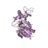
| Entry | Database: PDB / ID: 6qkv | |||||||||
|---|---|---|---|---|---|---|---|---|---|---|
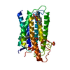
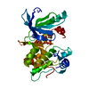
| Title | Structure of YibK from P. aeruginosa | |||||||||
 Components Components | tRNA (cytidine(34)-2'-O)-methyltransferase | |||||||||
 Keywords Keywords | TRANSFERASE / methyltransferase | |||||||||
| Function / homology |  Function and homology information Function and homology informationwobble position cytosine ribose methylation / wobble position uridine ribose methylation / tRNA (cytidine(34)-2'-O-ribose)-methyltransferase activity / tRNA (5-carboxymethylaminomethyluridine(34)-2'-O-ribose)-methyltransferase activity / tRNA (cytidine34-2'-O)-methyltransferase / tRNA methyltransferase activity / S-adenosylmethionine-dependent methyltransferase activity / RNA binding / identical protein binding / cytoplasm Similarity search - Function | |||||||||
| Biological species |   Pseudomonas aeruginosa (bacteria) Pseudomonas aeruginosa (bacteria) | |||||||||
| Method |  X-RAY DIFFRACTION / SYNCHROTRON / MOLECULAR REPLACEMENT / Resolution: 2.01 Å X-RAY DIFFRACTION / SYNCHROTRON / MOLECULAR REPLACEMENT / Resolution: 2.01 Å | |||||||||
 Authors Authors | Mikula, K.M. / Tascon, I. / Iwai, H. | |||||||||
| Funding support |  Finland, 2items Finland, 2items
| |||||||||
 Citation Citation | Journal: Front Chem / Year: 2021 Title: Tying up the Loose Ends: A Mathematically Knotted Protein. Authors: Hsu, S.D. / Lee, Y.C. / Mikula, K.M. / Backlund, S.M. / Tascon, I. / Goldman, A. / Iwai, H. | |||||||||
| History |
|
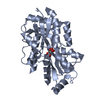
- Structure visualization
| Structure viewer | Molecule: MolmilJmol/JSmol |
|---|
- Downloads & links
-Download
| PDBx/mmCIF format | 6qkv.cif.gz | 75.8 KB | Display | PDBx/mmCIF format |
|---|---|---|---|---|
| PDB format | pdb6qkv.ent.gz | 56.2 KB | Display | PDB format |
| PDBx/mmJSON format | 6qkv.json.gz | Tree view | PDBx/mmJSON format | |
| Others |  Other downloads Other downloads |
-Validation report
| Arichive directory | https://data.pdbj.org/pub/pdb/validation_reports/qk/6qkvftp://data.pdbj.org/pub/pdb/validation_reports/qk/6qkv | HTTPS FTP |
|---|
-Related structure data
| Related structure data | 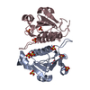 6qh8C 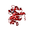 1mxiS S: Starting model for refinement C: citing same article ( |
|---|---|
| Similar structure data |


-Links
 PDBj
PDBj- Assembly
| Deposited unit | 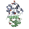
| ||||||||
|---|---|---|---|---|---|---|---|---|---|
| 1 |
| ||||||||
| Unit cell |
|
-Components
| #1: Protein | Mass: 17706.234 Da / Num. of mol.: 2 Source method: isolated from a genetically manipulated source Source: (gene. exp.) Pseudomonas aeruginosa (bacteria)Gene: trmL, C0044_37955, C8257_31370, DZ962_17365, PAMH19_2880 Production host: References: UniProt: A0A071LCY6, UniProt: Q9HU57*PLUS, tRNA (cytidine34-2'-O)-methyltransferase #2: Chemical | ChemComp-SO4 /   Mass: 96.063 Da / Num. of mol.: 6 / Source method: obtained synthetically / Formula: SO4 Mass: 96.063 Da / Num. of mol.: 6 / Source method: obtained synthetically / Formula: SO4#3: Chemical | ChemComp-GOL /   Mass: 92.094 Da / Num. of mol.: 5 / Source method: obtained synthetically / Formula: C3H8O3 Mass: 92.094 Da / Num. of mol.: 5 / Source method: obtained synthetically / Formula: C3H8O3#4: Water | ChemComp-HOH / |  Mass: 18.015 Da / Num. of mol.: 155 / Source method: isolated from a natural source / Formula: H2O Mass: 18.015 Da / Num. of mol.: 155 / Source method: isolated from a natural source / Formula: H2O |
|---|
-Experimental details
-Experiment
| Experiment | Method: X-RAY DIFFRACTION / Number of used crystals: 1 |
|---|
- Sample preparation
| Crystal | Density Matthews: 2.47 Å3/Da / Density % sol: 50.25 % |
|---|---|
| Crystal grow | Temperature: 293 K / Method: vapor diffusion, sitting drop Details: 0.15 M ammonium sulfate, 0.9 M lithium sulfate, 0.1 M sodium citrate buffer, pH 5.6 |
-Data collection
| Diffraction | Mean temperature: 100 K / Serial crystal experiment: N |
|---|---|
| Diffraction source | Source: SYNCHROTRON / Site: ESRF  / Beamline: ID14-4 / Wavelength: 0.9795 Å / Beamline: ID14-4 / Wavelength: 0.9795 Å |
| Detector | Type: ADSC QUANTUM 315 / Detector: CCD / Date: Jun 14, 2012 |
| Radiation | Protocol: SINGLE WAVELENGTH / Monochromatic (M) / Laue (L): M / Scattering type: x-ray |
| Radiation wavelength | Wavelength: 0.9795 Å / Relative weight: 1 |
| Reflection | Resolution: 2.01→46.7 Å / Num. obs: 23919 / % possible obs: 99.5 % / Redundancy: 5.8 % / Rrim(I) all: 0.071 / Net I/σ(I): 18.52 |
| Reflection shell | Resolution: 2.01→2.13 Å / Redundancy: 4.9 % / Mean I/σ(I) obs: 2.84 / Num. unique obs: 3813 / Rrim(I) all: 0.665 / % possible all: 99.2 |
- Processing
| Software |
| |||||||||||||||||||||||||||||||||||||||||||||||||||||||||||||||||||||||||||||||||||||||||||||||||||||||||
|---|---|---|---|---|---|---|---|---|---|---|---|---|---|---|---|---|---|---|---|---|---|---|---|---|---|---|---|---|---|---|---|---|---|---|---|---|---|---|---|---|---|---|---|---|---|---|---|---|---|---|---|---|---|---|---|---|---|---|---|---|---|---|---|---|---|---|---|---|---|---|---|---|---|---|---|---|---|---|---|---|---|---|---|---|---|---|---|---|---|---|---|---|---|---|---|---|---|---|---|---|---|---|---|---|---|---|
| Refinement | Method to determine structure: MOLECULAR REPLACEMENT Starting model: 1mxi Resolution: 2.01→46.699 Å / SU ML: 0.21 / Cross valid method: THROUGHOUT / σ(F): 1.35 / Phase error: 22.45
| |||||||||||||||||||||||||||||||||||||||||||||||||||||||||||||||||||||||||||||||||||||||||||||||||||||||||
| Solvent computation | Shrinkage radii: 0.9 Å / VDW probe radii: 1.11 Å | |||||||||||||||||||||||||||||||||||||||||||||||||||||||||||||||||||||||||||||||||||||||||||||||||||||||||
| Refinement step | Cycle: LAST / Resolution: 2.01→46.699 Å
| |||||||||||||||||||||||||||||||||||||||||||||||||||||||||||||||||||||||||||||||||||||||||||||||||||||||||
| Refine LS restraints |
| |||||||||||||||||||||||||||||||||||||||||||||||||||||||||||||||||||||||||||||||||||||||||||||||||||||||||
| LS refinement shell |
|
