 Movie
Movie Controller

[English] 日本語
 Yorodumi
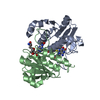
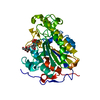
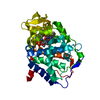
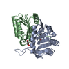
- PDB-1mxi: Structure of YibK from Haemophilus influenzae (HI0766): a Methylt... -
+ Open data

- Basic information
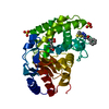
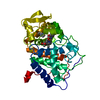
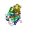
| Entry | Database: PDB / ID: 1mxi | ||||||
|---|---|---|---|---|---|---|---|
| Title | Structure of YibK from Haemophilus influenzae (HI0766): a Methyltransferase with a Cofactor Bound at a Site Formed by a Knot | ||||||
 Components Components | Hypothetical tRNA/rRNA methyltransferase HI0766 | ||||||
 Keywords Keywords | TRANSFERASE / methyltransferase / S-adenosylhomocysteine / spoU family / Structure 2 Function Project / S2F / Structural Genomics | ||||||
| Function / homology |  Function and homology information Function and homology informationwobble position cytosine ribose methylation / wobble position uridine ribose methylation / tRNA (cytidine(34)-2'-O-ribose)-methyltransferase activity / tRNA (5-carboxymethylaminomethyluridine(34)-2'-O-ribose)-methyltransferase activity / tRNA (cytidine34-2'-O)-methyltransferase / RNA binding / identical protein binding / cytoplasm Similarity search - Function | ||||||
| Biological species |  Haemophilus influenzae (bacteria) Haemophilus influenzae (bacteria) | ||||||
| Method |  X-RAY DIFFRACTION / SYNCHROTRON / MOLECULAR REPLACEMENT / Resolution: 1.7 Å X-RAY DIFFRACTION / SYNCHROTRON / MOLECULAR REPLACEMENT / Resolution: 1.7 Å | ||||||
 Authors Authors | Lim, K. / Zhang, H. / Tempczyk, A. / Bonander, N. / Toedt, J. / Howard, A. / Eisenstein, E. / Herzberg, O. / Structure 2 Function Project (S2F) | ||||||
 Citation Citation | Journal: Proteins / Year: 2003 Title: Structure of the YibK methyltransferase from Haemophilus influenzae (HI0766): a Cofactor Bound at a Site Formed by a Knot Authors: Lim, K. / Zhang, H. / Tempczyk, A. / Krajewski, W. / Bonander, N. / Toedt, J. / Howard, A. / Eisenstein, E. / Herzberg, O. | ||||||
| History |
|
- Structure visualization
| Structure viewer | Molecule: MolmilJmol/JSmol |
|---|
- Downloads & links
-Download
| PDBx/mmCIF format | 1mxi.cif.gz | 49.7 KB | Display | PDBx/mmCIF format |
|---|---|---|---|---|
| PDB format | pdb1mxi.ent.gz | 34.9 KB | Display | PDB format |
| PDBx/mmJSON format | 1mxi.json.gz | Tree view | PDBx/mmJSON format | |
| Others |  Other downloads Other downloads |
-Validation report
| Arichive directory | https://data.pdbj.org/pub/pdb/validation_reports/mx/1mxiftp://data.pdbj.org/pub/pdb/validation_reports/mx/1mxi | HTTPS FTP |
|---|
-Related structure data
| Related structure data |  1j85SC S: Starting model for refinement C: citing same article ( |
|---|---|
| Similar structure data | |
| Other databases |


-Links
 PDBj
PDBj- Assembly
| Deposited unit | 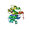
| ||||||||
|---|---|---|---|---|---|---|---|---|---|
| 1 | 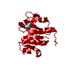
| ||||||||
| Unit cell |
|
-Components
| #1: Protein | Mass: 18427.414 Da / Num. of mol.: 1 Source method: isolated from a genetically manipulated source Source: (gene. exp.) Haemophilus influenzae (bacteria) / Gene: HI0766 / Plasmid: PET17B / Species (production host): Escherichia coli / Production host: References: UniProt: P44868, Transferases; Transferring one-carbon groups; Methyltransferases |
|---|---|
| #2: Chemical | ChemComp-IOD /   Mass: 126.904 Da / Num. of mol.: 1 / Source method: obtained synthetically / Formula: I Mass: 126.904 Da / Num. of mol.: 1 / Source method: obtained synthetically / Formula: I |
| #3: Chemical | ChemComp-SAH /   Type: L-peptide linking / Mass: 384.411 Da / Num. of mol.: 1 / Source method: obtained synthetically / Formula: C14H20N6O5S Type: L-peptide linking / Mass: 384.411 Da / Num. of mol.: 1 / Source method: obtained synthetically / Formula: C14H20N6O5S |
| #4: Water | ChemComp-HOH /  Mass: 18.015 Da / Num. of mol.: 153 / Source method: isolated from a natural source / Formula: H2O Mass: 18.015 Da / Num. of mol.: 153 / Source method: isolated from a natural source / Formula: H2O |
-Experimental details
-Experiment
| Experiment | Method: X-RAY DIFFRACTION / Number of used crystals: 1 |
|---|
- Sample preparation
| Crystal | Density Matthews: 1.87 Å3/Da / Density % sol: 34.27 % | ||||||||||||||||||||||||||||||||||||||||||||||||||||||
|---|---|---|---|---|---|---|---|---|---|---|---|---|---|---|---|---|---|---|---|---|---|---|---|---|---|---|---|---|---|---|---|---|---|---|---|---|---|---|---|---|---|---|---|---|---|---|---|---|---|---|---|---|---|---|---|
| Crystal grow | Temperature: 298 K Method: vapor diffusion, hanging drop, soaking of crystal with cofactor sah pH: 4.6 Details: 20% PMME 2000, 0.1 M sodium acetate, 0.2 M ammonium acetate, 3 % ethylene glycol, pH 4.6, Vapor diffusion, hanging drop, soaking of crystal with cofactor SAH, temperature 298K | ||||||||||||||||||||||||||||||||||||||||||||||||||||||
| Crystal grow | *PLUS pH: 7.5 / Method: vapor diffusion, hanging drop | ||||||||||||||||||||||||||||||||||||||||||||||||||||||
| Components of the solutions | *PLUS
|
-Data collection
| Diffraction | Mean temperature: 100 K |
|---|---|
| Diffraction source | Source: SYNCHROTRON / Site: APS  / Beamline: 17-ID / Wavelength: 1 Å / Beamline: 17-ID / Wavelength: 1 Å |
| Detector | Type: MARRESEARCH / Detector: CCD / Date: Oct 1, 2001 |
| Radiation | Protocol: SINGLE WAVELENGTH / Monochromatic (M) / Laue (L): M / Scattering type: x-ray |
| Radiation wavelength | Wavelength: 1 Å / Relative weight: 1 |
| Reflection | Resolution: 1.7→50 Å / Num. all: 15394 / Num. obs: 15394 / % possible obs: 93.7 % / Observed criterion σ(F): 0 / Observed criterion σ(I): 0 / Rmerge(I) obs: 0.04 / Net I/σ(I): 18.5 |
| Reflection shell | Resolution: 1.7→1.78 Å / Rmerge(I) obs: 0.223 / % possible all: 69.2 |
| Reflection | *PLUS Lowest resolution: 50 Å / Num. measured all: 281315 / Rmerge(I) obs: 0.04 |
| Reflection shell | *PLUS Highest resolution: 1.7 Å / % possible obs: 69.2 % |
- Processing
| Software |
| ||||||||||||||||
|---|---|---|---|---|---|---|---|---|---|---|---|---|---|---|---|---|---|
| Refinement | Method to determine structure: MOLECULAR REPLACEMENT Starting model: PDB 1j85 Resolution: 1.7→20 Å / Isotropic thermal model: isotropic / Cross valid method: THROUGHOUT / σ(F): 2 / Stereochemistry target values: Engh & Huber
| ||||||||||||||||
| Displacement parameters | Biso mean: 29 Å2 | ||||||||||||||||
| Refinement step | Cycle: LAST / Resolution: 1.7→20 Å
| ||||||||||||||||
| Refine LS restraints |
| ||||||||||||||||
| LS refinement shell | Resolution: 1.7→1.78 Å / Rfactor Rfree: 0.273 / Rfactor Rwork: 0.215 |
