 Movie
Movie Controller

[English] 日本語
 Yorodumi
- PDB-7bw1: Crystal structure of Steroid 5-alpha-reductase 2 in complex with ... -
+ Open data

- Basic information
| Entry | Database: PDB / ID: 7bw1 | |||||||||
|---|---|---|---|---|---|---|---|---|---|---|
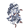
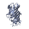
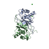
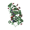
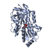
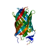
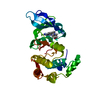
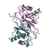
| Title | Crystal structure of Steroid 5-alpha-reductase 2 in complex with Finasteride | |||||||||
 Components Components | 3-oxo-5-alpha-steroid 4-dehydrogenase 2 | |||||||||
 Keywords Keywords | OXIDOREDUCTASE / Integral membrane protein / Reductase / Steroid | |||||||||
| Function / homology |  Function and homology information Function and homology information3-oxo-5alpha-steroid 4-dehydrogenase (NADP+) / biphenyl metabolic process / : / 3-oxo-5-alpha-steroid 4-dehydrogenase (NADP+) activity / 3-oxo-5-alpha-steroid 4-dehydrogenase activity / dibenzo-p-dioxin metabolic process / phthalate metabolic process / female genitalia development / : / response to follicle-stimulating hormone ...3-oxo-5alpha-steroid 4-dehydrogenase (NADP+) / biphenyl metabolic process / : / 3-oxo-5-alpha-steroid 4-dehydrogenase (NADP+) activity / 3-oxo-5-alpha-steroid 4-dehydrogenase activity / dibenzo-p-dioxin metabolic process / phthalate metabolic process / female genitalia development / : / response to follicle-stimulating hormone / Androgen biosynthesis / androgen biosynthetic process / testosterone biosynthetic process / cell body fiber / steroid biosynthetic process / steroid catabolic process / male genitalia development / response to steroid hormone / response to testosterone / hypothalamus development / androgen metabolic process / hippocampus development / response to nutrient levels / bone development / male gonad development / response to peptide hormone / cell-cell signaling / cell differentiation / response to xenobiotic stimulus / neuronal cell body / endoplasmic reticulum membrane Similarity search - Function | |||||||||
| Biological species |  Homo sapiens (human) Homo sapiens (human) | |||||||||
| Method |  X-RAY DIFFRACTION / SYNCHROTRON / MOLECULAR REPLACEMENT / Resolution: 2.8 Å X-RAY DIFFRACTION / SYNCHROTRON / MOLECULAR REPLACEMENT / Resolution: 2.8 Å | |||||||||
 Authors Authors | Xiao, Q. / Zhang, C. / Wei, Z. | |||||||||
| Funding support |  China, 2items China, 2items
| |||||||||
 Citation Citation | Journal: Res Sq / Year: 2020 Title: Structure of human steroid 5 alpha-reductase 2 with anti-androgen drug finasteride. Authors: Xiao, Q. / Wang, L. / Supekar, S. / Shen, T. / Liu, H. / Ye, F. / Huang, J. / Fan, H. / Wei, Z. / Zhang, C. | |||||||||
| History |
|
- Structure visualization
| Structure viewer | Molecule: MolmilJmol/JSmol |
|---|
- Downloads & links
-Download
| PDBx/mmCIF format | 7bw1.cif.gz | 117.5 KB | Display | PDBx/mmCIF format |
|---|---|---|---|---|
| PDB format | pdb7bw1.ent.gz | 89.2 KB | Display | PDB format |
| PDBx/mmJSON format | 7bw1.json.gz | Tree view | PDBx/mmJSON format | |
| Others |  Other downloads Other downloads |
-Validation report
| Arichive directory | https://data.pdbj.org/pub/pdb/validation_reports/bw/7bw1ftp://data.pdbj.org/pub/pdb/validation_reports/bw/7bw1 | HTTPS FTP |
|---|
-Related structure data
| Similar structure data |
|---|


-Links
 PDBj
PDBj


- Assembly
| Deposited unit | 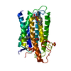
| ||||||||
|---|---|---|---|---|---|---|---|---|---|
| 1 |
| ||||||||
| Unit cell |
|
-Components
| #1: Protein | Mass: 28693.520 Da / Num. of mol.: 1 Source method: isolated from a genetically manipulated source Source: (gene. exp.) Homo sapiens (human) / Gene: SRD5A2 / Production host:  Baculovirus expression vector pFastBac1-HM Baculovirus expression vector pFastBac1-HMReferences: UniProt: P31213, 3-oxo-5alpha-steroid 4-dehydrogenase (NADP+) | ||||
|---|---|---|---|---|---|
| #2: Chemical | ChemComp-OLC / (  Mass: 356.540 Da / Num. of mol.: 1 / Source method: obtained synthetically / Formula: C21H40O4 Mass: 356.540 Da / Num. of mol.: 1 / Source method: obtained synthetically / Formula: C21H40O4 | ||||
| #3: Chemical | ChemComp-NDX / [[(  Mass: 1117.965 Da / Num. of mol.: 1 / Source method: obtained synthetically / Formula: C44H66N9O19P3 / Feature type: SUBJECT OF INVESTIGATION Mass: 1117.965 Da / Num. of mol.: 1 / Source method: obtained synthetically / Formula: C44H66N9O19P3 / Feature type: SUBJECT OF INVESTIGATION | ||||
| #4: Chemical |   Mass: 96.063 Da / Num. of mol.: 2 / Source method: obtained synthetically / Formula: SO4 Mass: 96.063 Da / Num. of mol.: 2 / Source method: obtained synthetically / Formula: SO4Has ligand of interest | Y | Has protein modification | Y | |
-Experimental details
-Experiment
| Experiment | Method: X-RAY DIFFRACTION / Number of used crystals: 1 |
|---|
- Sample preparation
| Crystal | Density Matthews: 3 Å3/Da / Density % sol: 59.02 % |
|---|---|
| Crystal grow | Temperature: 293 K / Method: lipidic cubic phase Details: 30% v/v PEG600, 100mM tris-sodium citrate ph5.0, 100mM sodium chloride, 100mM Lithium sulfate |
-Data collection
| Diffraction | Mean temperature: 100 K / Serial crystal experiment: N | |||||||||||||||||||||||||||||||||||||||||||||||||||||||||||||||||||||||||||||||||||||||||||||||||||||||||||||||||||||||||||||||||||||||||||||||||||||||||||||||||||||||||||||||||||||||||||||
|---|---|---|---|---|---|---|---|---|---|---|---|---|---|---|---|---|---|---|---|---|---|---|---|---|---|---|---|---|---|---|---|---|---|---|---|---|---|---|---|---|---|---|---|---|---|---|---|---|---|---|---|---|---|---|---|---|---|---|---|---|---|---|---|---|---|---|---|---|---|---|---|---|---|---|---|---|---|---|---|---|---|---|---|---|---|---|---|---|---|---|---|---|---|---|---|---|---|---|---|---|---|---|---|---|---|---|---|---|---|---|---|---|---|---|---|---|---|---|---|---|---|---|---|---|---|---|---|---|---|---|---|---|---|---|---|---|---|---|---|---|---|---|---|---|---|---|---|---|---|---|---|---|---|---|---|---|---|---|---|---|---|---|---|---|---|---|---|---|---|---|---|---|---|---|---|---|---|---|---|---|---|---|---|---|---|---|---|---|---|---|
| Diffraction source | Source: SYNCHROTRON / Site: APS  / Beamline: 23-ID-D / Wavelength: 0.979 Å / Beamline: 23-ID-D / Wavelength: 0.979 Å | |||||||||||||||||||||||||||||||||||||||||||||||||||||||||||||||||||||||||||||||||||||||||||||||||||||||||||||||||||||||||||||||||||||||||||||||||||||||||||||||||||||||||||||||||||||||||||||
| Detector | Type: DECTRIS PILATUS3 6M / Detector: PIXEL / Date: Aug 21, 2019 | |||||||||||||||||||||||||||||||||||||||||||||||||||||||||||||||||||||||||||||||||||||||||||||||||||||||||||||||||||||||||||||||||||||||||||||||||||||||||||||||||||||||||||||||||||||||||||||
| Radiation | Protocol: SINGLE WAVELENGTH / Monochromatic (M) / Laue (L): M / Scattering type: x-ray | |||||||||||||||||||||||||||||||||||||||||||||||||||||||||||||||||||||||||||||||||||||||||||||||||||||||||||||||||||||||||||||||||||||||||||||||||||||||||||||||||||||||||||||||||||||||||||||
| Radiation wavelength | Wavelength: 0.979 Å / Relative weight: 1 | |||||||||||||||||||||||||||||||||||||||||||||||||||||||||||||||||||||||||||||||||||||||||||||||||||||||||||||||||||||||||||||||||||||||||||||||||||||||||||||||||||||||||||||||||||||||||||||
| Reflection | Resolution: 2.8→40 Å / Num. obs: 9161 / % possible obs: 99.7 % / Redundancy: 9.6 % / Biso Wilson estimate: 68.94 Å2 / Rmerge(I) obs: 0.248 / Rpim(I) all: 0.079 / Rrim(I) all: 0.261 / Χ2: 1.131 / Net I/σ(I): 3.7 | |||||||||||||||||||||||||||||||||||||||||||||||||||||||||||||||||||||||||||||||||||||||||||||||||||||||||||||||||||||||||||||||||||||||||||||||||||||||||||||||||||||||||||||||||||||||||||||
| Reflection shell | Diffraction-ID: 1
|
- Processing
| Software |
| ||||||||||||||||||||||||||||||||||||||||||||||||||||||||||||||||||||||||||||||||||||||||||||||||||||
|---|---|---|---|---|---|---|---|---|---|---|---|---|---|---|---|---|---|---|---|---|---|---|---|---|---|---|---|---|---|---|---|---|---|---|---|---|---|---|---|---|---|---|---|---|---|---|---|---|---|---|---|---|---|---|---|---|---|---|---|---|---|---|---|---|---|---|---|---|---|---|---|---|---|---|---|---|---|---|---|---|---|---|---|---|---|---|---|---|---|---|---|---|---|---|---|---|---|---|---|---|---|
| Refinement | Method to determine structure: MOLECULAR REPLACEMENT / Resolution: 2.8→35.171 Å / SU ML: 0.39 / Cross valid method: THROUGHOUT / σ(F): 1.36 / Phase error: 31.2
| ||||||||||||||||||||||||||||||||||||||||||||||||||||||||||||||||||||||||||||||||||||||||||||||||||||
| Solvent computation | Shrinkage radii: 0.9 Å / VDW probe radii: 1.11 Å | ||||||||||||||||||||||||||||||||||||||||||||||||||||||||||||||||||||||||||||||||||||||||||||||||||||
| Displacement parameters | Biso max: 188.8 Å2 / Biso mean: 75.84 Å2 / Biso min: 39.23 Å2 | ||||||||||||||||||||||||||||||||||||||||||||||||||||||||||||||||||||||||||||||||||||||||||||||||||||
| Refinement step | Cycle: final / Resolution: 2.8→35.171 Å
| ||||||||||||||||||||||||||||||||||||||||||||||||||||||||||||||||||||||||||||||||||||||||||||||||||||
| Refine LS restraints |
| ||||||||||||||||||||||||||||||||||||||||||||||||||||||||||||||||||||||||||||||||||||||||||||||||||||
| LS refinement shell | Refine-ID: X-RAY DIFFRACTION / Rfactor Rfree error: 0
| ||||||||||||||||||||||||||||||||||||||||||||||||||||||||||||||||||||||||||||||||||||||||||||||||||||
| Refinement TLS params. | Method: refined / Refine-ID: X-RAY DIFFRACTION
| ||||||||||||||||||||||||||||||||||||||||||||||||||||||||||||||||||||||||||||||||||||||||||||||||||||
| Refinement TLS group |
|
