 Movie
Movie Controller


+ Open data

- Basic information
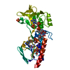
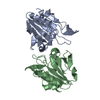
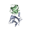
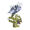
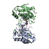
| Entry | Database: PDB / ID: 6qa0 | ||||||
|---|---|---|---|---|---|---|---|
| Title | MSRB3 - AA 1-137 | ||||||
 Components Components | Methionine-R-sulfoxide reductase B3 | ||||||
 Keywords Keywords | OXIDOREDUCTASE / Enzyme / Methionine Sulfoxide | ||||||
| Function / homology |  Function and homology information Function and homology informationL-methionine (R)-S-oxide reductase / L-methionine (R)-S-oxide reductase activity / peptide-methionine (R)-S-oxide reductase / peptide-methionine (R)-S-oxide reductase activity / Protein repair / protein repair / response to oxidative stress / endoplasmic reticulum / mitochondrion / zinc ion binding ...L-methionine (R)-S-oxide reductase / L-methionine (R)-S-oxide reductase activity / peptide-methionine (R)-S-oxide reductase / peptide-methionine (R)-S-oxide reductase activity / Protein repair / protein repair / response to oxidative stress / endoplasmic reticulum / mitochondrion / zinc ion binding / cytoplasm / cytosol Similarity search - Function | ||||||
| Biological species |  Homo sapiens (human) Homo sapiens (human) | ||||||
| Method |  X-RAY DIFFRACTION / SYNCHROTRON / MOLECULAR REPLACEMENT / Resolution: 1.709 Å X-RAY DIFFRACTION / SYNCHROTRON / MOLECULAR REPLACEMENT / Resolution: 1.709 Å | ||||||
 Authors Authors | Javitt, G. / Fass, D. | ||||||
 Citation Citation | Journal: Antioxid.Redox Signal. / Year: 2020 Title: Structure and Electron-Transfer Pathway of the Human Methionine Sulfoxide Reductase MsrB3. Authors: Javitt, G. / Cao, Z. / Resnick, E. / Gabizon, R. / Bulleid, N.J. / Fass, D. | ||||||
| History |
|
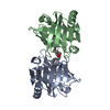
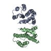
- Structure visualization
| Structure viewer | Molecule: MolmilJmol/JSmol |
|---|
- Downloads & links
-Download
| PDBx/mmCIF format | 6qa0.cif.gz | 80 KB | Display | PDBx/mmCIF format |
|---|---|---|---|---|
| PDB format | pdb6qa0.ent.gz | 57.8 KB | Display | PDB format |
| PDBx/mmJSON format | 6qa0.json.gz | Tree view | PDBx/mmJSON format | |
| Others |  Other downloads Other downloads |
-Validation report
| Summary document | 6qa0_validation.pdf.gz | 443.3 KB | Display | wwPDB validaton report |
|---|---|---|---|---|
| Full document | 6qa0_full_validation.pdf.gz | 443.5 KB | Display | |
| Data in XML | 6qa0_validation.xml.gz | 18.8 KB | Display | |
| Data in CIF | 6qa0_validation.cif.gz | 26.4 KB | Display | |
| Arichive directory | https://data.pdbj.org/pub/pdb/validation_reports/qa/6qa0ftp://data.pdbj.org/pub/pdb/validation_reports/qa/6qa0 | HTTPS FTP |
-Related structure data
| Related structure data |  6q9vC  3cezS S: Starting model for refinement C: citing same article ( |
|---|---|
| Similar structure data |


-Links
 PDBj
PDBj
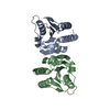
- Assembly
| Deposited unit | 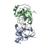
| ||||||||
|---|---|---|---|---|---|---|---|---|---|
| 1 |
| ||||||||
| Unit cell |
|
-Components
| #1: Protein | Mass: 15921.589 Da / Num. of mol.: 2 Source method: isolated from a genetically manipulated source Source: (gene. exp.) Homo sapiens (human) / Gene: MSRB3, UNQ1965/PRO4487 / Cell line (production host): HEK 293F / Production host: Homo sapiens (human)References: UniProt: Q8IXL7, peptide-methionine (R)-S-oxide reductase, L-methionine (R)-S-oxide reductase #2: Chemical | ChemComp-SO4 / |   Mass: 96.063 Da / Num. of mol.: 1 / Source method: obtained synthetically / Formula: SO4 Mass: 96.063 Da / Num. of mol.: 1 / Source method: obtained synthetically / Formula: SO4#3: Chemical | ChemComp-CL / |   Mass: 35.453 Da / Num. of mol.: 1 / Source method: obtained synthetically / Formula: Cl Mass: 35.453 Da / Num. of mol.: 1 / Source method: obtained synthetically / Formula: Cl#4: Chemical |   Mass: 65.409 Da / Num. of mol.: 2 / Source method: obtained synthetically / Formula: Zn Mass: 65.409 Da / Num. of mol.: 2 / Source method: obtained synthetically / Formula: Zn#5: Water | ChemComp-HOH / |  Mass: 18.015 Da / Num. of mol.: 334 / Source method: isolated from a natural source / Formula: H2O Mass: 18.015 Da / Num. of mol.: 334 / Source method: isolated from a natural source / Formula: H2OHas protein modification | Y | |
|---|
-Experimental details
-Experiment
| Experiment | Method: X-RAY DIFFRACTION / Number of used crystals: 1 |
|---|
- Sample preparation
| Crystal | Density Matthews: 2.68 Å3/Da / Density % sol: 54.19 % |
|---|---|
| Crystal grow | Temperature: 293 K / Method: vapor diffusion, hanging drop Details: 200mM NaCl, 100mM Sodium Cacodylate pH 7.4, 1.6M Ammonium Sulfate, 20% Glycerol |
-Data collection
| Diffraction | Mean temperature: 100 K / Serial crystal experiment: N | ||||||||||||||||||||||||
|---|---|---|---|---|---|---|---|---|---|---|---|---|---|---|---|---|---|---|---|---|---|---|---|---|---|
| Diffraction source | Source: SYNCHROTRON / Site: ESRF  / Beamline: ID23-2 / Wavelength: 0.87313 Å / Beamline: ID23-2 / Wavelength: 0.87313 Å | ||||||||||||||||||||||||
| Detector | Type: DECTRIS PILATUS3 X 2M / Detector: PIXEL / Date: Nov 29, 2018 | ||||||||||||||||||||||||
| Radiation | Protocol: SINGLE WAVELENGTH / Monochromatic (M) / Laue (L): M / Scattering type: x-ray | ||||||||||||||||||||||||
| Radiation wavelength | Wavelength: 0.87313 Å / Relative weight: 1 | ||||||||||||||||||||||||
| Reflection | Resolution: 1.71→42.86 Å / Num. obs: 37570 / % possible obs: 99.2 % / Redundancy: 4.2 % / Biso Wilson estimate: 24.88 Å2 / Rpim(I) all: 0.041 / Rrim(I) all: 0.084 / Net I/σ(I): 11.3 / Num. measured all: 159023 | ||||||||||||||||||||||||
| Reflection shell | Diffraction-ID: 1
|
- Processing
| Software |
| ||||||||||||||||||||||||||||||||||||||||||||||||||||||||||||||||||||||||||||||||||||||||||||||||||
|---|---|---|---|---|---|---|---|---|---|---|---|---|---|---|---|---|---|---|---|---|---|---|---|---|---|---|---|---|---|---|---|---|---|---|---|---|---|---|---|---|---|---|---|---|---|---|---|---|---|---|---|---|---|---|---|---|---|---|---|---|---|---|---|---|---|---|---|---|---|---|---|---|---|---|---|---|---|---|---|---|---|---|---|---|---|---|---|---|---|---|---|---|---|---|---|---|---|---|---|
| Refinement | Method to determine structure: MOLECULAR REPLACEMENT Starting model: 3CEZ Resolution: 1.709→42.649 Å / SU ML: 0.19 / Cross valid method: THROUGHOUT / σ(F): 1.33 / Phase error: 21.43 / Stereochemistry target values: ML
| ||||||||||||||||||||||||||||||||||||||||||||||||||||||||||||||||||||||||||||||||||||||||||||||||||
| Solvent computation | Shrinkage radii: 0.9 Å / VDW probe radii: 1.11 Å / Solvent model: FLAT BULK SOLVENT MODEL | ||||||||||||||||||||||||||||||||||||||||||||||||||||||||||||||||||||||||||||||||||||||||||||||||||
| Displacement parameters | Biso max: 92.1 Å2 / Biso mean: 30.8458 Å2 / Biso min: 14.6 Å2 | ||||||||||||||||||||||||||||||||||||||||||||||||||||||||||||||||||||||||||||||||||||||||||||||||||
| Refinement step | Cycle: final / Resolution: 1.709→42.649 Å
| ||||||||||||||||||||||||||||||||||||||||||||||||||||||||||||||||||||||||||||||||||||||||||||||||||
| LS refinement shell | Refine-ID: X-RAY DIFFRACTION / Rfactor Rfree error: 0 / Total num. of bins used: 13
|
