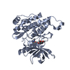
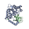
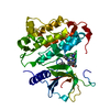
HYDROLASE / Glycoside hydrolase family 18 / complex N-glycans
Function / homology
Glycoside hydrolase family 18, BT1044-like / Glycoside hydrolase family 18, BT1044-like / Glycosyl hydrolases family 18 (GH18) active site / Glycosyl hydrolases family 18 (GH18) active site signature. / hydrolase activity, hydrolyzing O-glycosyl compounds / Prokaryotic membrane lipoprotein lipid attachment site profile. / Glycoside hydrolase superfamily / carbohydrate metabolic process / Endo-beta-N-acetylglucosaminidase family protein
Function and homology information
Biological species
Bacteroides thetaiotaomicron (bacteria)
Method
X-RAY DIFFRACTION / SYNCHROTRON / Resolution: 2.4 Å
Protocol: SINGLE WAVELENGTH / Monochromatic (M) / Laue (L): M / Scattering type: x-ray
Radiation wavelength
Wavelength: 0.9163 Å / Relative weight: 1
Reflection
Resolution: 2.4→48.5 Å / Num. obs: 27124 / % possible obs: 99.8 % / Redundancy: 11.1 % / CC1/2: 0.999 / Net I/σ(I): 12.6
Reflection shell
Resolution: 2.4→2.49 Å / Redundancy: 10.9 % / Mean I/σ(I) obs: 1.3 / Num. unique obs: 2797 / CC1/2: 0.835 / % possible all: 99.5
-
Processing
Software
Name
Classification
XDS
datareduction
XDS
datascaling
Aimless
datascaling
REFMAC
refinement
Coot
modelbuilding
Refinement
Resolution: 2.4→43.99 Å / Cor.coef. Fo:Fc: 0.969 / Cor.coef. Fo:Fc free: 0.95 / SU B: 28.885 / SU ML: 0.249 / Cross valid method: THROUGHOUT / ESU R: 0.2 / ESU R Free: 0.19 / Details: HYDROGENS HAVE BEEN ADDED IN THE RIDING POSITIONS
Rfactor
Num. reflection
% reflection
Selection details
Rfree
0.24364
1318
4.9 %
RANDOM
Rwork
0.19712
-
-
-
obs
0.1993
25784
99.81 %
-
Solvent computation
Ion probe radii: 0.8 Å / Shrinkage radii: 0.8 Å / VDW probe radii: 1.2 Å
 Movie
Movie Controller


 Open data
 Basic information
 Components
 Keywords
 Function and homology information
 Bacteroides thetaiotaomicron (bacteria)
 X-RAY DIFFRACTION /
  Authors
 United Kingdom, 1items
  Citation
 Structure visualization
 Downloads & links
 Other downloads
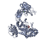


 PDBj
PDBj


 Assembly
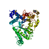
 Sample preparation
 Processing