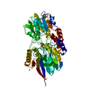
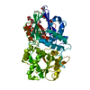
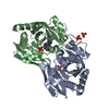
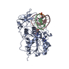
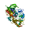
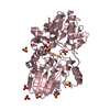
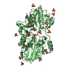
Entry Database : PDB / ID : 6q55Title Crystal structure of Cryptosporidium hominis CPSF3 in complex with Compound 61 Cleavage and Polyadenylation Specificity Factor 3 (CPSF3) Keywords / / / / Function / homology Function Domain/homology Component
/ / / / / / / / / / / / / / / / / / / / / / / / / / / / / / / / Biological species Cryptosporidium hominis TU502 (eukaryote)Method / / / Resolution : 2 Å Authors Palencia, A. / Swale, C. Funding support Organization Grant number Country European Commission iNEXT Grant 653706 French National Research Agency ANR-11-LABX- 0024 Parafrap
Journal : Sci Transl Med / Year : 2019Title : Metal-captured inhibition of pre-mRNA processing activity by CPSF3 controls Cryptosporidium infection.Authors : Swale, C. / Bougdour, A. / Gnahoui-David, A. / Tottey, J. / Georgeault, S. / Laurent, F. / Palencia, A. / Hakimi, M.A. History Deposition Dec 7, 2018 Deposition site / Processing site Revision 1.0 Nov 20, 2019 Provider / Type Revision 1.1 Sep 30, 2020 Group / Derived calculations / Category / pdbx_struct_conn_angle / struct_connItem _citation.title / _pdbx_struct_conn_angle.ptnr1_auth_seq_id ... _citation.title / _pdbx_struct_conn_angle.ptnr1_auth_seq_id / _pdbx_struct_conn_angle.ptnr3_auth_seq_id / _pdbx_struct_conn_angle.value / _struct_conn.pdbx_dist_value / _struct_conn.ptnr2_auth_seq_id Revision 1.2 Jan 24, 2024 Group / Database references / Refinement descriptionCategory chem_comp_atom / chem_comp_bond ... chem_comp_atom / chem_comp_bond / database_2 / pdbx_initial_refinement_model Item / _database_2.pdbx_database_accession
Show all Show less
 Movie
Movie Controller

 Yorodumi
 Open data
 Basic information
 Components
 Keywords
 Function and homology information
 Cryptosporidium hominis TU502 (eukaryote)
 X-RAY DIFFRACTION /
  Authors
 France, 2items
  Citation
 Structure visualization
 Downloads & links
 Other downloads

 PDBj
PDBj


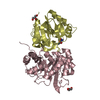
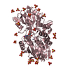
 Assembly


 Mass: 65.409 Da / Num. of mol.: 2 / Source method: obtained synthetically / Formula: Zn
 Mass: 223.010 Da / Num. of mol.: 1 / Source method: obtained synthetically / Formula: C10H12BO5 / Feature type: SUBJECT OF INVESTIGATION
 Mass: 60.095 Da / Num. of mol.: 4 / Source method: obtained synthetically / Formula: C3H8O / Comment: alkaloid*YM
 Mass: 92.094 Da / Num. of mol.: 5 / Source method: obtained synthetically / Formula: C3H8O3
 Mass: 24.305 Da / Num. of mol.: 2 / Source method: obtained synthetically / Formula: Mg
 Sample preparation
 Processing