 Movie
Movie Controller

[English] 日本語
 Yorodumi
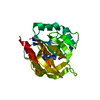
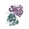
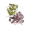
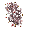
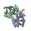
- PDB-3zr4: STRUCTURAL EVIDENCE FOR AMMONIA TUNNELING ACROSS THE (BETA-ALPHA)... -
+ Open data

- Basic information
| Entry | Database: PDB / ID: 3zr4 | ||||||
|---|---|---|---|---|---|---|---|
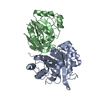
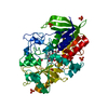
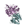
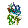
| Title | STRUCTURAL EVIDENCE FOR AMMONIA TUNNELING ACROSS THE (BETA-ALPHA)8 BARREL OF THE IMIDAZOLE GLYCEROL PHOSPHATE SYNTHASE BIENZYME COMPLEX | ||||||
 Components Components |
| ||||||
 Keywords Keywords | TRANSFERASE | ||||||
| Function / homology |  Function and homology information Function and homology informationimidazoleglycerol-phosphate synthase complex / imidazole glycerol-phosphate synthase / imidazoleglycerol-phosphate synthase activity / glutaminase / L-histidine biosynthetic process / glutaminase activity / lyase activity / cytoplasm Similarity search - Function | ||||||
| Biological species |   THERMOTOGA MARITIMA (bacteria) THERMOTOGA MARITIMA (bacteria) | ||||||
| Method |  X-RAY DIFFRACTION / SYNCHROTRON / MOLECULAR REPLACEMENT / Resolution: 2.41 Å X-RAY DIFFRACTION / SYNCHROTRON / MOLECULAR REPLACEMENT / Resolution: 2.41 Å | ||||||
 Authors Authors | Vega, M.C. / Kuper, J. / Haeger, M.C. / Mohrlueder, J. / Marquardt, S. / Sterner, R. / Wilmanns, M. | ||||||
 Citation Citation | Journal: Chem.Biol. / Year: 2012 Title: Catalysis Uncoupling in a Glutamine Amidotransferase Bienzyme by Unblocking the Glutaminase Active Site. Authors: List, F. / Vega, M.C. / Razeto, A. / Hager, M.C. / Sterner, R. / Wilmanns, M. #1: Journal: Structure / Year: 2002Title: Structural Evidence for Ammonia Tunneling Across the (Beta Alpha)(8) Barrel of the Imidazole Glycerol Phosphate Synthase Bienzyme Complex. Authors: Douangamath, A. / Walker, M. / Beismann-Driemeyer, S. / Vega-Fernandez, M.C. / Sterner, R. / Wilmanns, M. | ||||||
| History |
| ||||||
| Remark 700 | SHEET THE SHEET STRUCTURE OF THIS MOLECULE IS BIFURCATED. IN ORDER TO REPRESENT THIS FEATURE IN ... SHEET THE SHEET STRUCTURE OF THIS MOLECULE IS BIFURCATED. IN ORDER TO REPRESENT THIS FEATURE IN THE SHEET RECORDS BELOW, TWO SHEETS ARE DEFINED. |
- Structure visualization
| Structure viewer | Molecule: MolmilJmol/JSmol |
|---|
- Downloads & links
-Download
| PDBx/mmCIF format | 3zr4.cif.gz | 524.9 KB | Display | PDBx/mmCIF format |
|---|---|---|---|---|
| PDB format | pdb3zr4.ent.gz | 436.7 KB | Display | PDB format |
| PDBx/mmJSON format | 3zr4.json.gz | Tree view | PDBx/mmJSON format | |
| Others |  Other downloads Other downloads |
-Validation report
| Arichive directory | https://data.pdbj.org/pub/pdb/validation_reports/zr/3zr4ftp://data.pdbj.org/pub/pdb/validation_reports/zr/3zr4 | HTTPS FTP |
|---|
-Related structure data
| Related structure data |  2wjzC  1gpwS S: Starting model for refinement C: citing same article ( |
|---|---|
| Similar structure data |


-Links
 PDBj
PDBj


- Assembly
| Deposited unit | 
| ||||||||||||||||||||
|---|---|---|---|---|---|---|---|---|---|---|---|---|---|---|---|---|---|---|---|---|---|
| 1 | 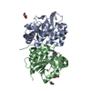
| ||||||||||||||||||||
| 2 | 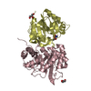
| ||||||||||||||||||||
| 3 | 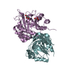
| ||||||||||||||||||||
| Unit cell |
| ||||||||||||||||||||
| Noncrystallographic symmetry (NCS) | NCS oper:
|
-Components
| #1: Protein | Mass: 27753.871 Da / Num. of mol.: 3 Source method: isolated from a genetically manipulated source Source: (gene. exp.) THERMOTOGA MARITIMA (bacteria) / Plasmid: RBSIISPHI-TMHISF / Production host: References: UniProt: Q9X0C6, Lyases; Carbon-carbon lyases; Oxo-acid-lyases #2: Protein | Mass: 23130.564 Da / Num. of mol.: 3 Source method: isolated from a genetically manipulated source Source: (gene. exp.) THERMOTOGA MARITIMA (bacteria) / Plasmid: RBSIISPHI-TMHISH / Production host: References: UniProt: Q9X0C8, Transferases; Glycosyltransferases; Pentosyltransferases #3: Chemical | ChemComp-GOL /   Mass: 92.094 Da / Num. of mol.: 7 / Source method: obtained synthetically / Formula: C3H8O3 Mass: 92.094 Da / Num. of mol.: 7 / Source method: obtained synthetically / Formula: C3H8O3#4: Chemical |   Type: L-peptide linking / Mass: 146.144 Da / Num. of mol.: 2 / Source method: obtained synthetically / Formula: C5H10N2O3 Type: L-peptide linking / Mass: 146.144 Da / Num. of mol.: 2 / Source method: obtained synthetically / Formula: C5H10N2O3#5: Water | ChemComp-HOH / |  Mass: 18.015 Da / Num. of mol.: 204 / Source method: isolated from a natural source / Formula: H2O Mass: 18.015 Da / Num. of mol.: 204 / Source method: isolated from a natural source / Formula: H2O |
|---|
-Experimental details
-Experiment
| Experiment | Method: X-RAY DIFFRACTION / Number of used crystals: 1 |
|---|
- Sample preparation
| Crystal | Density Matthews: 2.35 Å3/Da / Density % sol: 47.6 % / Description: NONE |
|---|
-Data collection
| Diffraction | Mean temperature: 100 K | |||||||||||||||
|---|---|---|---|---|---|---|---|---|---|---|---|---|---|---|---|---|
| Diffraction source | Source: SYNCHROTRON / Site: EMBL/DESY, HAMBURG  / Beamline: X13 / Wavelength: 0.802 / Beamline: X13 / Wavelength: 0.802 | |||||||||||||||
| Detector | Type: MARRESEARCH / Detector: CCD | |||||||||||||||
| Radiation | Protocol: SINGLE WAVELENGTH / Monochromatic (M) / Laue (L): M / Scattering type: x-ray | |||||||||||||||
| Radiation wavelength | Wavelength: 0.802 Å / Relative weight: 1 | |||||||||||||||
| Reflection twin |
| |||||||||||||||
| Reflection | Resolution: 2.4→55.95 Å / Num. obs: 51685 / % possible obs: 95.5 % / Observed criterion σ(I): -3 / Redundancy: 2.4 % / Rmerge(I) obs: 0.05 / Net I/σ(I): 16.8 | |||||||||||||||
| Reflection shell | Resolution: 2.4→2.5 Å / Redundancy: 2.3 % / Rmerge(I) obs: 0.34 / Mean I/σ(I) obs: 2.6 / % possible all: 88.9 |
- Processing
| Software |
| ||||||||||||||||||||||||||||||||||||||||||||||||||||||||||||||||||||||||||||||||||||||||||||||||||||||||||||||||||||||||||||||||||||||||||||||||||||||||||||||||||||||||||||||||||||||
|---|---|---|---|---|---|---|---|---|---|---|---|---|---|---|---|---|---|---|---|---|---|---|---|---|---|---|---|---|---|---|---|---|---|---|---|---|---|---|---|---|---|---|---|---|---|---|---|---|---|---|---|---|---|---|---|---|---|---|---|---|---|---|---|---|---|---|---|---|---|---|---|---|---|---|---|---|---|---|---|---|---|---|---|---|---|---|---|---|---|---|---|---|---|---|---|---|---|---|---|---|---|---|---|---|---|---|---|---|---|---|---|---|---|---|---|---|---|---|---|---|---|---|---|---|---|---|---|---|---|---|---|---|---|---|---|---|---|---|---|---|---|---|---|---|---|---|---|---|---|---|---|---|---|---|---|---|---|---|---|---|---|---|---|---|---|---|---|---|---|---|---|---|---|---|---|---|---|---|---|---|---|---|---|
| Refinement | Method to determine structure: MOLECULAR REPLACEMENT Starting model: PDB ENTRY 1GPW Resolution: 2.41→55.96 Å / Cor.coef. Fo:Fc: 0.948 / Cor.coef. Fo:Fc free: 0.907 / SU B: 16.587 / SU ML: 0.171 / Cross valid method: THROUGHOUT / ESU R: 0.157 / ESU R Free: 0.065 / Stereochemistry target values: MAXIMUM LIKELIHOOD / Details: HYDROGENS HAVE BEEN ADDED IN THE RIDING POSITIONS.
| ||||||||||||||||||||||||||||||||||||||||||||||||||||||||||||||||||||||||||||||||||||||||||||||||||||||||||||||||||||||||||||||||||||||||||||||||||||||||||||||||||||||||||||||||||||||
| Solvent computation | Ion probe radii: 0.8 Å / Shrinkage radii: 0.8 Å / VDW probe radii: 1.4 Å / Solvent model: MASK | ||||||||||||||||||||||||||||||||||||||||||||||||||||||||||||||||||||||||||||||||||||||||||||||||||||||||||||||||||||||||||||||||||||||||||||||||||||||||||||||||||||||||||||||||||||||
| Displacement parameters | Biso mean: 43.957 Å2
| ||||||||||||||||||||||||||||||||||||||||||||||||||||||||||||||||||||||||||||||||||||||||||||||||||||||||||||||||||||||||||||||||||||||||||||||||||||||||||||||||||||||||||||||||||||||
| Refinement step | Cycle: LAST / Resolution: 2.41→55.96 Å
| ||||||||||||||||||||||||||||||||||||||||||||||||||||||||||||||||||||||||||||||||||||||||||||||||||||||||||||||||||||||||||||||||||||||||||||||||||||||||||||||||||||||||||||||||||||||
| Refine LS restraints |
|