 Movie
Movie Controller

[English] 日本語
 Yorodumi
- PDB-6pzk: Cryo-EM Structure of the Respiratory Syncytial Virus Polymerase (... -
+ Open data

- Basic information
| Entry | Database: PDB / ID: 6pzk | ||||||
|---|---|---|---|---|---|---|---|
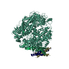
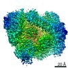
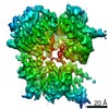
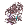
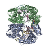
| Title | Cryo-EM Structure of the Respiratory Syncytial Virus Polymerase (L) Protein Bound by the Tetrameric Phosphoprotein (P) | ||||||
 Components Components |
| ||||||
 Keywords Keywords | VIRAL PROTEIN / RNA-binding protein / RSV / RdRp / RNA-dependent RNA polymerase / PRNTase / polyribonucleotidyl transferase / RNA capping / viral replication | ||||||
| Function / homology |  Function and homology information Function and homology informationRespiratory syncytial virus genome transcription / Translation of respiratory syncytial virus mRNAs / GDP polyribonucleotidyltransferase / negative stranded viral RNA replication / Respiratory syncytial virus genome replication / RSV-host interactions / Assembly and release of respiratory syncytial virus (RSV) virions / Maturation of hRSV A proteins / Respiratory syncytial virus (RSV) attachment and entry / Hydrolases; Acting on acid anhydrides; In phosphorus-containing anhydrides ...Respiratory syncytial virus genome transcription / Translation of respiratory syncytial virus mRNAs / GDP polyribonucleotidyltransferase / negative stranded viral RNA replication / Respiratory syncytial virus genome replication / RSV-host interactions / Assembly and release of respiratory syncytial virus (RSV) virions / Maturation of hRSV A proteins / Respiratory syncytial virus (RSV) attachment and entry / Hydrolases; Acting on acid anhydrides; In phosphorus-containing anhydrides / viral life cycle / virion component / symbiont-mediated suppression of host NF-kappaB cascade / host cell cytoplasm / mRNA 5'-cap (guanine-N7-)-methyltransferase activity / RNA-directed RNA polymerase / RNA-directed RNA polymerase activity / GTPase activity / ATP binding / metal ion binding Similarity search - Function | ||||||
| Biological species |  Human respiratory syncytial virus A2 Human respiratory syncytial virus A2 | ||||||
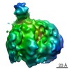
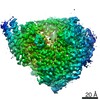
| Method | ELECTRON MICROSCOPY / single particle reconstruction / cryo EM / Resolution: 3.2 Å | ||||||
 Authors Authors | Gilman, M.S.A. / McLellan, J.S. | ||||||
 Citation Citation | Journal: Cell / Year: 2019 Title: Structure of the Respiratory Syncytial Virus Polymerase Complex. Authors: Morgan S A Gilman / Cheng Liu / Amy Fung / Ishani Behera / Paul Jordan / Peter Rigaux / Nina Ysebaert / Sergey Tcherniuk / Julien Sourimant / Jean-François Eléouët / Priscila Sutto-Ortiz ...Authors: Morgan S A Gilman / Cheng Liu / Amy Fung / Ishani Behera / Paul Jordan / Peter Rigaux / Nina Ysebaert / Sergey Tcherniuk / Julien Sourimant / Jean-François Eléouët / Priscila Sutto-Ortiz / Etienne Decroly / Dirk Roymans / Zhinan Jin / Jason S McLellan /    Abstract: Numerous interventions are in clinical development for respiratory syncytial virus (RSV) infection, including small molecules that target viral transcription and replication. These processes are ...Numerous interventions are in clinical development for respiratory syncytial virus (RSV) infection, including small molecules that target viral transcription and replication. These processes are catalyzed by a complex comprising the RNA-dependent RNA polymerase (L) and the tetrameric phosphoprotein (P). RSV P recruits multiple proteins to the polymerase complex and, with the exception of its oligomerization domain, is thought to be intrinsically disordered. Despite their critical roles in RSV transcription and replication, structures of L and P have remained elusive. Here, we describe the 3.2-Å cryo-EM structure of RSV L bound to tetrameric P. The structure reveals a striking tentacular arrangement of P, with each of the four monomers adopting a distinct conformation. The structure also rationalizes inhibitor escape mutants and mutations observed in live-attenuated vaccine candidates. These results provide a framework for determining the molecular underpinnings of RSV replication and transcription and should facilitate the design of effective RSV inhibitors. | ||||||
| History |
|
- Structure visualization
| Movie |
Movie viewer |
|---|---|
| Structure viewer | Molecule: MolmilJmol/JSmol |
- Downloads & links
-Download
| PDBx/mmCIF format | 6pzk.cif.gz | 328.3 KB | Display | PDBx/mmCIF format |
|---|---|---|---|---|
| PDB format | pdb6pzk.ent.gz | 244.7 KB | Display | PDB format |
| PDBx/mmJSON format | 6pzk.json.gz | Tree view | PDBx/mmJSON format | |
| Others |  Other downloads Other downloads |
-Validation report
| Arichive directory | https://data.pdbj.org/pub/pdb/validation_reports/pz/6pzkftp://data.pdbj.org/pub/pdb/validation_reports/pz/6pzk | HTTPS FTP |
|---|
-Related structure data
| Related structure data | 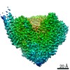 20536MC M: map data used to model this data C: citing same article ( |
|---|---|
| Similar structure data |

-Links
 PDBj
PDBj
- Assembly
| Deposited unit | 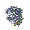
|
|---|---|
| 1 |
|
-Components
| #1: Protein | Mass: 254482.750 Da / Num. of mol.: 1 Source method: isolated from a genetically manipulated source Source: (gene. exp.) Human respiratory syncytial virus A2 / Strain: A2 / Cell line (production host): Sf9 / Production host:   Spodoptera frugiperda (fall armyworm) Spodoptera frugiperda (fall armyworm)References: UniProt: P28887, RNA-directed RNA polymerase, mRNA (guanine-N7)-methyltransferase, Transferases; Transferring phosphorus-containing groups; Nucleotidyltransferases, GDP polyribonucleotidyltransferase |
|---|---|
| #2: Protein | Mass: 29062.895 Da / Num. of mol.: 4 Source method: isolated from a genetically manipulated source Source: (gene. exp.) Human respiratory syncytial virus A2 / Strain: A2 / Cell line (production host): Sf9 / Production host: Spodoptera frugiperda (fall armyworm) / References: UniProt: P03421 |
-Experimental details
-Experiment
| Experiment | Method: ELECTRON MICROSCOPY |
|---|---|
| EM experiment | Aggregation state: PARTICLE / 3D reconstruction method: single particle reconstruction |
- Sample preparation
| Component | Name: Respiratory Syncytial Virus Polymerase (L) Protein Bound by the Tetrameric Phosphoprotein (P) Type: COMPLEX / Entity ID: all / Source: RECOMBINANT | |||||||||||||||||||||||||
|---|---|---|---|---|---|---|---|---|---|---|---|---|---|---|---|---|---|---|---|---|---|---|---|---|---|---|
| Molecular weight | Value: 0.37 MDa / Experimental value: NO | |||||||||||||||||||||||||
| Source (natural) | Organism: Human respiratory syncytial virus A2 | |||||||||||||||||||||||||
| Source (recombinant) | Organism: Spodoptera frugiperda (fall armyworm) | |||||||||||||||||||||||||
| Buffer solution | pH: 8 | |||||||||||||||||||||||||
| Buffer component |
| |||||||||||||||||||||||||
| Specimen | Conc.: 0.29 mg/ml / Embedding applied: NO / Shadowing applied: NO / Staining applied: NO / Vitrification applied: YES | |||||||||||||||||||||||||
| Specimen support | Grid material: GOLD | |||||||||||||||||||||||||
| Vitrification | Instrument: FEI VITROBOT MARK IV / Cryogen name: ETHANE |
- Electron microscopy imaging
| Experimental equipment |  Model: Titan Krios / Image courtesy: FEI Company |
|---|---|
| Microscopy | Model: FEI TITAN KRIOS |
| Electron gun | Electron source:  FIELD EMISSION GUN / Accelerating voltage: 300 kV / Illumination mode: FLOOD BEAM FIELD EMISSION GUN / Accelerating voltage: 300 kV / Illumination mode: FLOOD BEAM |
| Electron lens | Mode: BRIGHT FIELD |
| Image recording | Electron dose: 48 e/Å2 / Detector mode: COUNTING / Film or detector model: GATAN K2 SUMMIT (4k x 4k) |
- Processing
| EM software |
| |||||||||||||||
|---|---|---|---|---|---|---|---|---|---|---|---|---|---|---|---|---|
| CTF correction | Type: PHASE FLIPPING AND AMPLITUDE CORRECTION | |||||||||||||||
| Symmetry | Point symmetry: C1 (asymmetric) | |||||||||||||||
| 3D reconstruction | Resolution: 3.2 Å / Resolution method: FSC 0.143 CUT-OFF / Num. of particles: 196720 / Symmetry type: POINT |
