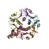
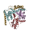
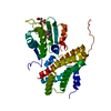
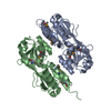
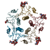
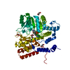
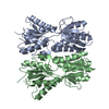
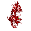
- PDB-6pzj: Structure of the N-terminal domain (residues 43-304) of Methyl-ac... -
+
データを開く
IDまたはキーワード:
読み込み中...
-
基本情報
登録情報
データベース: PDB / ID: 6pzj
タイトル
Structure of the N-terminal domain (residues 43-304) of Methyl-accepting chemotaxis protein from Leptospira interrogans serogroup Icterohaemorrhagiae serovar Copenhageni (strain Fiocruz L1-130)
要素
Methyl-accepting chemotaxis protein
キーワード
SIGNALING PROTEIN / SSGCID / Structural Genomics / Seattle Structural Genomics Center for Infectious Disease
温度: 290 K / 手法: 蒸気拡散法, シッティングドロップ法 / pH: 9.5 詳細: RigakuReagents JCSG+ screen A7: 20% (w/V) PEG 8000, 100mM CHES / NaOH pH 9.5: LpinA.18975.a.B2.PW38653 at 19.51mg/ml: cryo: 20% EG in 2 steps: tray 310977 a7: puck hqx5-3. For phasing: ...詳細: RigakuReagents JCSG+ screen A7: 20% (w/V) PEG 8000, 100mM CHES / NaOH pH 9.5: LpinA.18975.a.B2.PW38653 at 19.51mg/ml: cryo: 20% EG in 2 steps: tray 310977 a7: puck hqx5-3. For phasing: Microlytic MCSG1 screen condition H3: 20% (w/V) PEG 3350, 200mM Lithium acetate: LpinA.18975.a.B2.PW38653 at 19.51mg/ml. A crystal from this condition was soaked for 15sec in a mix of 90% reservoir and 10% 2.5M NaI in EG, and for another 15sec in a mix of 80% reservoir and 20% 2.5M NaI in EG, and flash frozen for in-house data collection: tray 310978 h3: puck hqx5-12
 ムービー
ムービー コントローラー


 データを開く
 基本情報
基本情報 要素
要素 キーワード
キーワード 機能・相同性情報
 Leptospira interrogans serogroup Icterohaemorrhagiae serovar copenhageni (バクテリア)
 X線回折 /
  データ登録者
 引用
引用 構造の表示
構造の表示 ダウンロードとリンク
 その他のダウンロード


 PDBj
PDBj


 集合体
集合体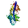

 分子量: 35.453 Da / 分子数: 1 / 由来タイプ: 合成 / 式: Cl
 分子量: 62.068 Da / 分子数: 2 / 由来タイプ: 合成 / 式: C2H6O2
 分子量: 18.015 Da / 分子数: 221 / 由来タイプ: 天然 / 式: H2O
 試料調製
試料調製
 解析
解析