 Movie
Movie Controller

[English] 日本語
 Yorodumi
- PDB-3huu: Crystal structure of transcription regulator like protein from St... -
+ Open data

- Basic information
| Entry | Database: PDB / ID: 3huu | ||||||
|---|---|---|---|---|---|---|---|
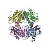
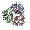
| Title | Crystal structure of transcription regulator like protein from Staphylococcus haemolyticus | ||||||
 Components Components | Transcription regulator like protein | ||||||
 Keywords Keywords | Transcription regulator / PSI-II / NYSGXRC / 11235m / Lac I / Structural Genomics / Protein Structure Initiative / New York SGX Research Center for Structural Genomics | ||||||
| Function / homology |  Function and homology information Function and homology informationtranscription cis-regulatory region binding / DNA-binding transcription factor activity Similarity search - Function | ||||||
| Biological species |  Staphylococcus haemolyticus (bacteria) Staphylococcus haemolyticus (bacteria) | ||||||
| Method |  X-RAY DIFFRACTION / SYNCHROTRON / SAD / Resolution: 1.95 Å X-RAY DIFFRACTION / SYNCHROTRON / SAD / Resolution: 1.95 Å | ||||||
 Authors Authors | Agarwal, R. / Burley, S.K. / Swaminathan, S. / New York SGX Research Center for Structural Genomics (NYSGXRC) | ||||||
 Citation Citation | Journal: To be Published Title: Crystal structure of transcription regulator like protein from Staphylococcus haemolyticus Authors: Agarwal, R. / Burley, S.K. / Swaminathan, S. | ||||||
| History |
|
- Structure visualization
| Structure viewer | Molecule: MolmilJmol/JSmol |
|---|
- Downloads & links
-Download
| PDBx/mmCIF format | 3huu.cif.gz | 214.2 KB | Display | PDBx/mmCIF format |
|---|---|---|---|---|
| PDB format | pdb3huu.ent.gz | 173.3 KB | Display | PDB format |
| PDBx/mmJSON format | 3huu.json.gz | Tree view | PDBx/mmJSON format | |
| Others |  Other downloads Other downloads |
-Validation report
| Arichive directory | https://data.pdbj.org/pub/pdb/validation_reports/hu/3huuftp://data.pdbj.org/pub/pdb/validation_reports/hu/3huu | HTTPS FTP |
|---|
-Related structure data
| Similar structure data | |
|---|---|
| Other databases |


-Links
 PDBj
PDBj
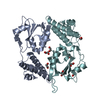
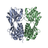
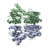
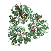
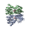
- Assembly
| Deposited unit | 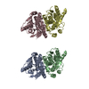
| ||||||||
|---|---|---|---|---|---|---|---|---|---|
| 1 | 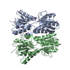
| ||||||||
| 2 | 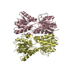
| ||||||||
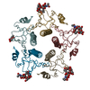
| Unit cell |
|
-Components
| #1: Protein | Mass: 34704.223 Da / Num. of mol.: 4 Source method: isolated from a genetically manipulated source Source: (gene. exp.) Staphylococcus haemolyticus (bacteria) / Strain: JCSC1435 / Gene: SH1407 / Plasmid: pSGX3(BC) / Production host: #2: Water | ChemComp-HOH / |  Mass: 18.015 Da / Num. of mol.: 128 / Source method: isolated from a natural source / Formula: H2O Mass: 18.015 Da / Num. of mol.: 128 / Source method: isolated from a natural source / Formula: H2O |
|---|
-Experimental details
-Experiment
| Experiment | Method: X-RAY DIFFRACTION / Number of used crystals: 1 |
|---|
- Sample preparation
| Crystal | Density Matthews: 2.36 Å3/Da / Density % sol: 47.95 % |
|---|---|
| Crystal grow | Temperature: 298 K / Method: vapor diffusion, sitting drop / pH: 7.5 Details: 10% PEG 8K, 8% Ethylene Glycol, 0.1M Hepes, pH 7.5, VAPOR DIFFUSION, SITTING DROP, temperature 298K |
-Data collection
| Diffraction | Mean temperature: 100 K |
|---|---|
| Diffraction source | Source: SYNCHROTRON / Site: NSLS  / Beamline: X29A / Wavelength: 0.9791 Å / Beamline: X29A / Wavelength: 0.9791 Å |
| Detector | Type: ADSC QUANTUM 315 / Detector: CCD / Date: Jun 6, 2009 / Details: mirrors |
| Radiation | Monochromator: Si-III / Protocol: SINGLE WAVELENGTH / Monochromatic (M) / Laue (L): M / Scattering type: x-ray |
| Radiation wavelength | Wavelength: 0.9791 Å / Relative weight: 1 |
| Reflection | Resolution: 1.95→50 Å / Num. all: 82791 / Num. obs: 82791 / % possible obs: 89.7 % / Observed criterion σ(F): 0 / Observed criterion σ(I): 0 / Redundancy: 7.5 % / Biso Wilson estimate: 16.6 Å2 / Rmerge(I) obs: 0.06 / Net I/σ(I): 10.5 |
| Reflection shell | Resolution: 1.95→1.98 Å / Redundancy: 7.1 % / Rmerge(I) obs: 0.43 / Mean I/σ(I) obs: 3.5 / Num. unique all: 4504 / % possible all: 96.8 |
- Processing
| Software |
| |||||||||||||||||||||||||
|---|---|---|---|---|---|---|---|---|---|---|---|---|---|---|---|---|---|---|---|---|---|---|---|---|---|---|
| Refinement | Method to determine structure: SAD Starting model: None Resolution: 1.95→43.76 Å / Rfactor Rfree error: 0.006 / Data cutoff high absF: 103598.74 / Data cutoff low absF: 0 / Isotropic thermal model: RESTRAINED / Cross valid method: THROUGHOUT / σ(F): 0 / σ(I): 0 / Stereochemistry target values: Engh & Huber Details: The missing residues and missing atoms are due to weak or no electron density.
| |||||||||||||||||||||||||
| Solvent computation | Solvent model: FLAT MODEL / Bsol: 38.5588 Å2 / ksol: 0.351416 e/Å3 | |||||||||||||||||||||||||
| Displacement parameters | Biso mean: 31.5 Å2
| |||||||||||||||||||||||||
| Refine analyze |
| |||||||||||||||||||||||||
| Refinement step | Cycle: LAST / Resolution: 1.95→43.76 Å
| |||||||||||||||||||||||||
| Refine LS restraints |
| |||||||||||||||||||||||||
| LS refinement shell | Resolution: 1.95→2.07 Å / Rfactor Rfree error: 0.016 / Total num. of bins used: 6
| |||||||||||||||||||||||||
| Xplor file |
|
