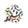
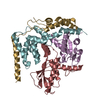
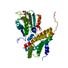
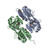
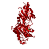
- PDB-6pzj: Structure of the N-terminal domain (residues 43-304) of Methyl-ac... -
+
Open data
ID or keywords:
Loading...
-
Basic information
Entry
Database: PDB / ID: 6pzj
Title
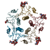
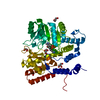
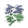
Structure of the N-terminal domain (residues 43-304) of Methyl-accepting chemotaxis protein from Leptospira interrogans serogroup Icterohaemorrhagiae serovar Copenhageni (strain Fiocruz L1-130)
Components
Methyl-accepting chemotaxis protein
Keywords
SIGNALING PROTEIN / SSGCID / Structural Genomics / Seattle Structural Genomics Center for Infectious Disease
Mass: 18.015 Da / Num. of mol.: 221 / Source method: isolated from a natural source / Formula: H2O
Has ligand of interest
N
-
Experimental details
-
Experiment
Experiment
Method: X-RAY DIFFRACTION / Number of used crystals: 1
-
Sample preparation
Crystal
Density Matthews: 2.4 Å3/Da / Density % sol: 48.8 %
Crystal grow
Temperature: 290 K / Method: vapor diffusion, sitting drop / pH: 9.5 Details: RigakuReagents JCSG+ screen A7: 20% (w/V) PEG 8000, 100mM CHES / NaOH pH 9.5: LpinA.18975.a.B2.PW38653 at 19.51mg/ml: cryo: 20% EG in 2 steps: tray 310977 a7: puck hqx5-3. For phasing: ...Details: RigakuReagents JCSG+ screen A7: 20% (w/V) PEG 8000, 100mM CHES / NaOH pH 9.5: LpinA.18975.a.B2.PW38653 at 19.51mg/ml: cryo: 20% EG in 2 steps: tray 310977 a7: puck hqx5-3. For phasing: Microlytic MCSG1 screen condition H3: 20% (w/V) PEG 3350, 200mM Lithium acetate: LpinA.18975.a.B2.PW38653 at 19.51mg/ml. A crystal from this condition was soaked for 15sec in a mix of 90% reservoir and 10% 2.5M NaI in EG, and for another 15sec in a mix of 80% reservoir and 20% 2.5M NaI in EG, and flash frozen for in-house data collection: tray 310978 h3: puck hqx5-12
In the structure databanks used in Yorodumi, some data are registered as the other names, "COVID-19 virus" and "2019-nCoV". Here are the details of the virus and the list of structure data.
Jan 31, 2019. EMDB accession codes are about to change! (news from PDBe EMDB page)
EMDB accession codes are about to change! (news from PDBe EMDB page)
The allocation of 4 digits for EMDB accession codes will soon come to an end. Whilst these codes will remain in use, new EMDB accession codes will include an additional digit and will expand incrementally as the available range of codes is exhausted. The current 4-digit format prefixed with “EMD-” (i.e. EMD-XXXX) will advance to a 5-digit format (i.e. EMD-XXXXX), and so on. It is currently estimated that the 4-digit codes will be depleted around Spring 2019, at which point the 5-digit format will come into force.
The EM Navigator/Yorodumi systems omit the EMD- prefix.
Related info.:Q: What is EMD? / ID/Accession-code notation in Yorodumi/EM Navigator
Yorodumi is a browser for structure data from EMDB, PDB, SASBDB, etc.
This page is also the successor to EM Navigator detail page, and also detail information page/front-end page for Omokage search.
The word "yorodu" (or yorozu) is an old Japanese word meaning "ten thousand". "mi" (miru) is to see.
Related info.:EMDB / PDB / SASBDB / Comparison of 3 databanks / Yorodumi Search / Aug 31, 2016. New EM Navigator & Yorodumi / Yorodumi Papers / Jmol/JSmol / Function and homology information / Changes in new EM Navigator and Yorodumi
 Movie
Movie Controller

 Yorodumi
 Open data
 Basic information
 Components
 Keywords
 Function and homology information
 Leptospira interrogans serogroup Icterohaemorrhagiae serovar copenhageni (bacteria)
 X-RAY DIFFRACTION /
  Authors
 Citation
 Structure visualization
 Downloads & links
 Other downloads


 PDBj
PDBj


 Assembly
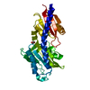

 Mass: 35.453 Da / Num. of mol.: 1 / Source method: obtained synthetically / Formula: Cl
 Mass: 62.068 Da / Num. of mol.: 2 / Source method: obtained synthetically / Formula: C2H6O2
 Mass: 18.015 Da / Num. of mol.: 221 / Source method: isolated from a natural source / Formula: H2O
 Sample preparation
 Processing