 Movie
Movie Controller

[English] 日本語
 Yorodumi
- PDB-6pwz: Crystal structure of human uridine-cytidine kinase 2 complexed wi... -
+ Open data

- Basic information
| Entry | Database: PDB / ID: 6pwz | ||||||
|---|---|---|---|---|---|---|---|
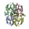
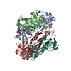
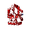
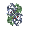
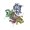
| Title | Crystal structure of human uridine-cytidine kinase 2 complexed with 2'-azidocytidine | ||||||
 Components Components | Uridine-cytidine kinase 2 | ||||||
 Keywords Keywords | TRANSFERASE/TRANSFERASE INHIBITOR / uridine cytidine kinase / nucleotide binding / TRANSFERASE-TRANSFERASE INHIBITOR complex | ||||||
| Function / homology |  Function and homology information Function and homology informationCTP salvage / uridine/cytidine kinase / uridine kinase activity / Pyrimidine salvage / cytidine kinase activity / UMP salvage / cellular response to oxygen levels / feeding behavior / response to axon injury / ATP binding ...CTP salvage / uridine/cytidine kinase / uridine kinase activity / Pyrimidine salvage / cytidine kinase activity / UMP salvage / cellular response to oxygen levels / feeding behavior / response to axon injury / ATP binding / identical protein binding / cytoplasm / cytosol Similarity search - Function | ||||||
| Biological species |  Homo sapiens (human) Homo sapiens (human) | ||||||
| Method |  X-RAY DIFFRACTION / SYNCHROTRON / MOLECULAR REPLACEMENT / Resolution: 3 Å X-RAY DIFFRACTION / SYNCHROTRON / MOLECULAR REPLACEMENT / Resolution: 3 Å | ||||||
 Authors Authors | Cuthbert, B.J. / Goulding, C.W. | ||||||
| Funding support |  United States, 1items United States, 1items
| ||||||
 Citation Citation | Journal: To Be Published Title: Incorporation of novel azido-nucleotides into RNA Authors: Nainar, S. / Cuthbert, B.J. / Goulding, C.W. / Spitale, R.C. | ||||||
| History |
|
- Structure visualization
| Structure viewer | Molecule: MolmilJmol/JSmol |
|---|
- Downloads & links
-Download
| PDBx/mmCIF format | 6pwz.cif.gz | 429 KB | Display | PDBx/mmCIF format |
|---|---|---|---|---|
| PDB format | pdb6pwz.ent.gz | 283.9 KB | Display | PDB format |
| PDBx/mmJSON format | 6pwz.json.gz | Tree view | PDBx/mmJSON format | |
| Others |  Other downloads Other downloads |
-Validation report
| Arichive directory | https://data.pdbj.org/pub/pdb/validation_reports/pw/6pwzftp://data.pdbj.org/pub/pdb/validation_reports/pw/6pwz | HTTPS FTP |
|---|
-Related structure data
| Related structure data |  6n55S S: Starting model for refinement |
|---|---|
| Similar structure data |


-Links
 PDBj
PDBj- Assembly
| Deposited unit | 
| ||||||||||||
|---|---|---|---|---|---|---|---|---|---|---|---|---|---|
| 1 | 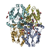
| ||||||||||||
| 2 | 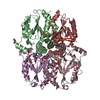
| ||||||||||||
| Unit cell |
|
-Components
| #1: Protein | Mass: 28108.830 Da / Num. of mol.: 8 Source method: isolated from a genetically manipulated source Source: (gene. exp.) Homo sapiens (human) / Gene: UCK2, UMPK / Production host:  #2: Chemical | ChemComp-PO4 /   Mass: 94.971 Da / Num. of mol.: 8 / Source method: obtained synthetically / Formula: PO4 Mass: 94.971 Da / Num. of mol.: 8 / Source method: obtained synthetically / Formula: PO4#3: Chemical | ChemComp-GOL /   Mass: 92.094 Da / Num. of mol.: 30 / Source method: obtained synthetically / Formula: C3H8O3 Mass: 92.094 Da / Num. of mol.: 30 / Source method: obtained synthetically / Formula: C3H8O3#4: Chemical |   Mass: 269.237 Da / Num. of mol.: 3 / Source method: isolated from a natural source / Formula: C9H13N6O4 / Feature type: SUBJECT OF INVESTIGATION Mass: 269.237 Da / Num. of mol.: 3 / Source method: isolated from a natural source / Formula: C9H13N6O4 / Feature type: SUBJECT OF INVESTIGATION#5: Water | ChemComp-HOH / |  Mass: 18.015 Da / Num. of mol.: 322 / Source method: isolated from a natural source / Formula: H2O Mass: 18.015 Da / Num. of mol.: 322 / Source method: isolated from a natural source / Formula: H2OHas ligand of interest | Y | |
|---|
-Experimental details
-Experiment
| Experiment | Method: X-RAY DIFFRACTION / Number of used crystals: 1 |
|---|
- Sample preparation
| Crystal | Density Matthews: 2.7 Å3/Da / Density % sol: 54.45 % |
|---|---|
| Crystal grow | Temperature: 298 K / Method: vapor diffusion, hanging drop / pH: 6.5 Details: 0.1 M Bis-Tris, pH 6.5, 25% pentaerythritol ethoxylate (15/4 EO/OH) |
-Data collection
| Diffraction | Mean temperature: 100 K / Serial crystal experiment: N |
|---|---|
| Diffraction source | Source: SYNCHROTRON / Site: ALS / Beamline: 5.0.2 / Wavelength: 1 Å |
| Detector | Type: DECTRIS PILATUS 6M / Detector: PIXEL / Date: Jun 5, 2018 |
| Radiation | Monochromator: double crystal Si(111) / Protocol: SINGLE WAVELENGTH / Monochromatic (M) / Laue (L): M / Scattering type: x-ray |
| Radiation wavelength | Wavelength: 1 Å / Relative weight: 1 |
| Reflection | Resolution: 3→83.2 Å / Num. obs: 48286 / % possible obs: 99.8 % / Redundancy: 6.1 % / Biso Wilson estimate: 69.63 Å2 / CC1/2: 0.996 / Rmerge(I) obs: 0.057 / Rpim(I) all: 0.057 / Rrim(I) all: 0.081 / Net I/σ(I): 6.5 |
| Reflection shell | Resolution: 3→3.11 Å / Redundancy: 2 % / Rmerge(I) obs: 0.432 / Mean I/σ(I) obs: 1.65 / Num. unique obs: 4783 / CC1/2: 0.619 / Rpim(I) all: 0.432 / Rrim(I) all: 0.611 / % possible all: 99.9 |
- Processing
| Software |
| |||||||||||||||||||||||||||||||||||||||||||||||||||||||||||||||
|---|---|---|---|---|---|---|---|---|---|---|---|---|---|---|---|---|---|---|---|---|---|---|---|---|---|---|---|---|---|---|---|---|---|---|---|---|---|---|---|---|---|---|---|---|---|---|---|---|---|---|---|---|---|---|---|---|---|---|---|---|---|---|---|---|
| Refinement | Method to determine structure: MOLECULAR REPLACEMENT Starting model: PDB entry 6N55 Resolution: 3→83.2 Å / SU ML: 0.3918 / Cross valid method: FREE R-VALUE / σ(F): 1.35 / Phase error: 25.985
| |||||||||||||||||||||||||||||||||||||||||||||||||||||||||||||||
| Solvent computation | Shrinkage radii: 0.9 Å / VDW probe radii: 1.11 Å | |||||||||||||||||||||||||||||||||||||||||||||||||||||||||||||||
| Displacement parameters | Biso mean: 62.58 Å2 | |||||||||||||||||||||||||||||||||||||||||||||||||||||||||||||||
| Refinement step | Cycle: LAST / Resolution: 3→83.2 Å
| |||||||||||||||||||||||||||||||||||||||||||||||||||||||||||||||
| Refine LS restraints |
| |||||||||||||||||||||||||||||||||||||||||||||||||||||||||||||||
| LS refinement shell |
|
