 Movie
Movie Controller


+ Open data

- Basic information
| Entry | Database: PDB / ID: 1ufq | ||||||
|---|---|---|---|---|---|---|---|
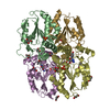
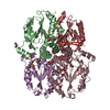
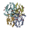
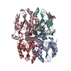
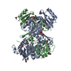
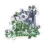
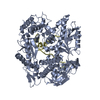
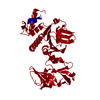
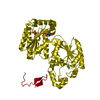
| Title | Crystal structure of ligand-free human uridine-cytidine kinase 2 | ||||||
 Components Components | Uridine-cytidine kinase 2 | ||||||
 Keywords Keywords | TRANSFERASE / ALPHA/BETA MONONUCLEOTIDE-BINDING HOLD | ||||||
| Function / homology |  Function and homology information Function and homology informationuridine/cytidine kinase / CTP salvage / uridine kinase activity / Pyrimidine salvage / cytidine kinase activity / UMP salvage / cellular response to oxygen levels / feeding behavior / response to axon injury / ATP binding ...uridine/cytidine kinase / CTP salvage / uridine kinase activity / Pyrimidine salvage / cytidine kinase activity / UMP salvage / cellular response to oxygen levels / feeding behavior / response to axon injury / ATP binding / identical protein binding / cytoplasm / cytosol Similarity search - Function | ||||||
| Biological species |  Homo sapiens (human) Homo sapiens (human) | ||||||
| Method |  X-RAY DIFFRACTION / SYNCHROTRON / MOLECULAR REPLACEMENT / Resolution: 2.5 Å X-RAY DIFFRACTION / SYNCHROTRON / MOLECULAR REPLACEMENT / Resolution: 2.5 Å | ||||||
 Authors Authors | Suzuki, N.N. / Koizumi, K. / Fukushima, M. / Matsuda, A. / Inagaki, F. | ||||||
 Citation Citation | Journal: STRUCTURE / Year: 2004 Title: Structural basis for the specificity, catalysis, and regulation of human uridine-cytidine kinase Authors: Suzuki, N.N. / Koizumi, K. / Fukushima, M. / Matsuda, A. / Inagaki, F. #1: Journal: ACTA CRYSTALLOGR.,SECT.D / Year: 2003 Title: Crystallization and preliminary X-ray analysis of human uridine-cytidine kinase 2 Authors: Suzuki, N.N. / Koizumi, K. / Fukushima, M. / Matsuda, A. / Inagaki, F. | ||||||
| History |
|
- Structure visualization
| Structure viewer | Molecule: MolmilJmol/JSmol |
|---|
- Downloads & links
-Download
| PDBx/mmCIF format | 1ufq.cif.gz | 173.8 KB | Display | PDBx/mmCIF format |
|---|---|---|---|---|
| PDB format | pdb1ufq.ent.gz | 137.5 KB | Display | PDB format |
| PDBx/mmJSON format | 1ufq.json.gz | Tree view | PDBx/mmJSON format | |
| Others |  Other downloads Other downloads |
-Validation report
| Arichive directory | https://data.pdbj.org/pub/pdb/validation_reports/uf/1ufqftp://data.pdbj.org/pub/pdb/validation_reports/uf/1ufq | HTTPS FTP |
|---|
-Related structure data
| Related structure data | 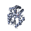 1udwSC 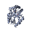 1ueiC 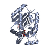 1uejC 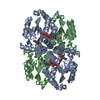 1uj2C S: Starting model for refinement C: citing same article ( |
|---|---|
| Similar structure data |

-Links
 PDBj
PDBj- Assembly
| Deposited unit | 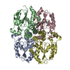
| ||||||||
|---|---|---|---|---|---|---|---|---|---|
| 1 |
| ||||||||
| 2 | 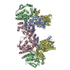
| ||||||||
| 3 | 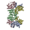
| ||||||||
| Unit cell |
|
-Components
| #1: Protein | Mass: 28262.994 Da / Num. of mol.: 4 / Fragment: residues 1-250 Source method: isolated from a genetically manipulated source Source: (gene. exp.) Homo sapiens (human) / Plasmid: pGEX-6P / Species (production host): Escherichia coli / Production host:  #2: Water | ChemComp-HOH / |  Mass: 18.015 Da / Num. of mol.: 130 / Source method: isolated from a natural source / Formula: H2O Mass: 18.015 Da / Num. of mol.: 130 / Source method: isolated from a natural source / Formula: H2O |
|---|
-Experimental details
-Experiment
| Experiment | Method: X-RAY DIFFRACTION / Number of used crystals: 1 |
|---|
- Sample preparation
| Crystal | Density Matthews: 3.35 Å3/Da / Density % sol: 62.98 % |
|---|---|
| Crystal grow | Temperature: 293 K / Method: vapor diffusion, sitting drop / pH: 6.9 Details: PEG400, PEG3350, glycerol, HEPES, pH 6.9, VAPOR DIFFUSION, SITTING DROP, temperature 293K |
-Data collection
| Diffraction | Mean temperature: 100 K |
|---|---|
| Diffraction source | Source: SYNCHROTRON / Site: SPring-8  / Beamline: BL44XU / Wavelength: 0.9 Å / Beamline: BL44XU / Wavelength: 0.9 Å |
| Detector | Type: OXFORD / Detector: CCD / Date: May 27, 2002 |
| Radiation | Monochromator: rotated-inclined monochromator, vertical focusing mirror Protocol: SINGLE WAVELENGTH / Monochromatic (M) / Laue (L): M / Scattering type: x-ray |
| Radiation wavelength | Wavelength: 0.9 Å / Relative weight: 1 |
| Reflection | Resolution: 2.5→40.41 Å / Num. all: 38404 / Num. obs: 38404 / % possible obs: 87.4 % / Observed criterion σ(F): 0 / Biso Wilson estimate: 21.5 Å2 / Rmerge(I) obs: 0.052 / Net I/σ(I): 12 |
| Reflection shell | Resolution: 2.5→2.59 Å / Rmerge(I) obs: 0.217 / Mean I/σ(I) obs: 3.2 / Num. unique all: 3910 / % possible all: 90.3 |
- Processing
| Software |
| ||||||||||||||||||||||||||||||||||||||||||||||||||||||||||||||||||||||||||||||||
|---|---|---|---|---|---|---|---|---|---|---|---|---|---|---|---|---|---|---|---|---|---|---|---|---|---|---|---|---|---|---|---|---|---|---|---|---|---|---|---|---|---|---|---|---|---|---|---|---|---|---|---|---|---|---|---|---|---|---|---|---|---|---|---|---|---|---|---|---|---|---|---|---|---|---|---|---|---|---|---|---|---|
| Refinement | Method to determine structure: MOLECULAR REPLACEMENT Starting model: PDB ENTRY 1UDW Resolution: 2.5→40.41 Å / Rfactor Rfree error: 0.005 / Data cutoff high absF: 302132.67 / Data cutoff low absF: 0 / Isotropic thermal model: RESTRAINED / Cross valid method: THROUGHOUT / σ(F): 0 / Stereochemistry target values: Engh & Huber
| ||||||||||||||||||||||||||||||||||||||||||||||||||||||||||||||||||||||||||||||||
| Solvent computation | Solvent model: FLAT MODEL / Bsol: 39.2919 Å2 / ksol: 0.344901 e/Å3 | ||||||||||||||||||||||||||||||||||||||||||||||||||||||||||||||||||||||||||||||||
| Displacement parameters | Biso mean: 41.1 Å2
| ||||||||||||||||||||||||||||||||||||||||||||||||||||||||||||||||||||||||||||||||
| Refine analyze |
| ||||||||||||||||||||||||||||||||||||||||||||||||||||||||||||||||||||||||||||||||
| Refinement step | Cycle: LAST / Resolution: 2.5→40.41 Å
| ||||||||||||||||||||||||||||||||||||||||||||||||||||||||||||||||||||||||||||||||
| Refine LS restraints |
| ||||||||||||||||||||||||||||||||||||||||||||||||||||||||||||||||||||||||||||||||
| LS refinement shell | Resolution: 2.5→2.66 Å / Rfactor Rfree error: 0.014 / Total num. of bins used: 6
| ||||||||||||||||||||||||||||||||||||||||||||||||||||||||||||||||||||||||||||||||
| Xplor file |
|
