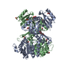
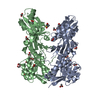
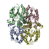
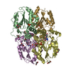
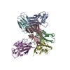
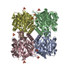
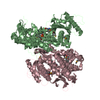
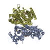
[English] 日本語
Yorodumi
- PDB-4pg6: Crystal structure of S. aureus Homoserine Dehydrogenase at pH7.0 -

+
Open data


ID or keywords:

Loading...

-
Basic information

Entry
Database: PDB / ID: 4pg6
TitleCrystal structure of S. aureus Homoserine Dehydrogenase at pH7.0
ComponentsHomoserine dehydrogenase
KeywordsOXIDOREDUCTASE / Aspartic acid pathway / pH sensitivity / hydride transfer / ACT domain
Function / homology
Function and homology information


homoserine dehydrogenase / homoserine dehydrogenase activity / threonine biosynthetic process / methionine biosynthetic process / NADP binding
Similarity search - Function
Alpha-Beta Plaits - #3100 / : / Homoserine dehydrogenase C-terminal domain / Homoserine dehydrogenase / Homoserine dehydrogenase, conserved site / Homoserine dehydrogenase signature. / Homoserine dehydrogenase, catalytic / Homoserine dehydrogenase / Aspartate/homoserine dehydrogenase, NAD-binding / Homoserine dehydrogenase, NAD binding domain ...Alpha-Beta Plaits - #3100 / : / Homoserine dehydrogenase C-terminal domain / Homoserine dehydrogenase / Homoserine dehydrogenase, conserved site / Homoserine dehydrogenase signature. / Homoserine dehydrogenase, catalytic / Homoserine dehydrogenase / Aspartate/homoserine dehydrogenase, NAD-binding / Homoserine dehydrogenase, NAD binding domain / Dihydrodipicolinate Reductase; domain 2 / Dihydrodipicolinate Reductase; domain 2 / NAD(P)-binding Rossmann-like Domain / Alpha-Beta Plaits / NAD(P)-binding domain superfamily / Rossmann fold / 2-Layer Sandwich / 3-Layer(aba) Sandwich / Alpha Beta
Similarity search - Domain/homology
ACETATE ION / DI(HYDROXYETHYL)ETHER / Homoserine dehydrogenase
Similarity search - Component
Biological speciesStaphylococcus aureus (bacteria)
MethodX-RAY DIFFRACTION / SYNCHROTRON / MOLECULAR REPLACEMENT / molecular replacement / Resolution: 2.2 Å
Model detailsStructure solved using the data obtained from the crystals grown at pH6.0 crystallization condition
AuthorsNavratna, V. / Gopal, B.
CitationJournal: Acta Crystallogr.,Sect.D / Year: 2015
Title: Structural basis for the catalytic mechanism of homoserine dehydrogenase.
Authors: Navratna, V. / Reddy, G. / Gopal, B.
History
DepositionMay 1, 2014Deposition site: RCSB / Processing site: RCSB
Revision 1.0May 13, 2015Provider: repository / Type: Initial release
Revision 1.1Jul 1, 2015Group: Database references
Revision 1.2Sep 27, 2017Group: Author supporting evidence / Derived calculations ...Author supporting evidence / Derived calculations / Refinement description / Source and taxonomy
Category: entity_src_gen / pdbx_audit_support ...entity_src_gen / pdbx_audit_support / pdbx_struct_oper_list / software
Item: _entity_src_gen.pdbx_alt_source_flag / _pdbx_struct_oper_list.symmetry_operation
Revision 1.3Sep 27, 2023Group: Data collection / Database references / Refinement description
Category: chem_comp_atom / chem_comp_bond ...chem_comp_atom / chem_comp_bond / database_2 / pdbx_initial_refinement_model / struct_ncs_dom_lim
Item: _database_2.pdbx_DOI / _database_2.pdbx_database_accession ..._database_2.pdbx_DOI / _database_2.pdbx_database_accession / _struct_ncs_dom_lim.beg_auth_comp_id / _struct_ncs_dom_lim.beg_label_asym_id / _struct_ncs_dom_lim.beg_label_comp_id / _struct_ncs_dom_lim.beg_label_seq_id / _struct_ncs_dom_lim.end_auth_comp_id / _struct_ncs_dom_lim.end_label_asym_id / _struct_ncs_dom_lim.end_label_comp_id / _struct_ncs_dom_lim.end_label_seq_id

-
Structure visualization

Structure viewerMolecule:
MolmilJmol/JSmol

Downloads & links

-
Assembly

Deposited unit
A: Homoserine dehydrogenase
B: Homoserine dehydrogenase
hetero molecules


Theoretical massNumber of molelcules
Total (without water)104,36524
Polymers102,7682
Non-polymers1,59722
Water4,738263
1


  • Idetical with deposited unit
  • defined by author&software
TypeNameSymmetry operationNumber
identity operation1_555x,y,z1
Buried area6210 Å2
ΔGint4 kcal/mol
Surface area36130 Å2
MethodPISA
Unit cell
Length a, b, c (Å)72.640, 116.880, 118.620
Angle α, β, γ (deg.)90.00, 90.00, 90.00
Int Tables number19
Space group name H-MP212121
Noncrystallographic symmetry (NCS)NCS domain:
IDEns-IDDetails
11A
21B

NCS domain segments:

Component-ID: _ / Ens-ID: 1 / End auth comp-ID: VAL / End label comp-ID: VAL / Refine code: _

Dom-IDBeg auth comp-IDBeg label comp-IDAuth asym-IDLabel asym-IDAuth seq-IDLabel seq-ID
1LYSLYSAA2 - 42622 - 446
2METMETBB1 - 42621 - 446

-
Components

-
Protein , 1 types, 2 molecules AB

#1: Protein Homoserine dehydrogenase


Mass: 51384.184 Da / Num. of mol.: 2
Source method: isolated from a genetically manipulated source
Source: (gene. exp.) Staphylococcus aureus (bacteria)
Gene: hom, CH51_06975, JN533_07120, QT38_06630, SASCBU26_01217
Plasmid: pET15b / Production host: Escherichia coli (E. coli) / Strain (production host): Rosetta (DE3) pLysS / References: UniProt: W8UB14, homoserine dehydrogenase

-
Non-polymers , 5 types, 285 molecules

#2: Chemical
ChemComp-ACT / ACETATE ION


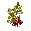
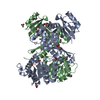
Mass: 59.044 Da / Num. of mol.: 13 / Source method: obtained synthetically / Formula: C2H3O2
#3: Chemical ChemComp-GOL / GLYCEROL / GLYCERIN / PROPANE-1,2,3-TRIOL


Mass: 92.094 Da / Num. of mol.: 3 / Source method: obtained synthetically / Formula: C3H8O3
#4: Chemical ChemComp-PEG / DI(HYDROXYETHYL)ETHER


Mass: 106.120 Da / Num. of mol.: 3 / Source method: obtained synthetically / Formula: C4H10O3
#5: Chemical ChemComp-DMS / DIMETHYL SULFOXIDE


Mass: 78.133 Da / Num. of mol.: 3 / Source method: obtained synthetically / Formula: C2H6OS / Comment: DMSO, precipitant*YM
#6: Water ChemComp-HOH / water


Mass: 18.015 Da / Num. of mol.: 263 / Source method: isolated from a natural source / Formula: H2O

-
Experimental details

-
Experiment

ExperimentMethod: X-RAY DIFFRACTION / Number of used crystals: 1

-
Sample preparation

CrystalDensity Matthews: 2.45 Å3/Da / Density % sol: 49.79 %
Crystal growTemperature: 293 K / Method: vapor diffusion, sitting drop / pH: 7
Details: 0.2M Magnesium acetate, 16% PEG3350, 0.1M HEPES, pH7.0, 5%Glycerol, 20% Iso-propanol

-
Data collection

DiffractionMean temperature: 100 K
Diffraction sourceSource: SYNCHROTRON / Site: ESRF / Beamline: BM14 / Wavelength: 0.97625 Å
DetectorType: MARMOSAIC 225 mm CCD / Detector: CCD / Date: Jul 26, 2012
RadiationProtocol: SINGLE WAVELENGTH / Monochromatic (M) / Laue (L): M / Scattering type: x-ray
Radiation wavelengthWavelength: 0.97625 Å / Relative weight: 1
ReflectionRedundancy: 8.3 % / Number: 433739 / Rsym value: 0.075 / D res high: 2.2 Å / D res low: 118.62 Å / Num. obs: 52050 / % possible obs: 100
Diffraction reflection shell
Highest resolution (Å)Lowest resolution (Å)IDRmerge(I) obsRsym valueRedundancy
6.9634.7310.0360.0367.6
4.926.9610.0370.0378.1
4.024.9210.0640.0648.3
3.484.0210.0670.0678.3
3.113.4810.070.078.4
2.843.1110.0870.0878.4
2.632.8410.1370.1378.4
2.462.6310.2060.2068.4
2.322.4610.3160.3168.4
2.22.3210.4420.4428.4
ReflectionResolution: 2.2→118.62 Å / Num. obs: 52050 / % possible obs: 100 % / Redundancy: 8.3 % / Biso Wilson estimate: 47.2 Å2 / Net I/σ(I): 17.3
Reflection shell

Diffraction-ID: 1 / Rejects: _

Resolution (Å)Redundancy (%)Rmerge(I) obsMean I/σ(I) obsNum. measured allNum. unique allRpim(I) allRsym valueNet I/σ(I) obs% possible all
2.2-2.328.40.4421.76287374940.1620.4424.8100
2.32-2.468.40.3162.45949770830.1160.3166.6100
2.46-2.638.40.2063.65628166980.0750.2069.6100
2.63-2.848.40.1375.45245362510.050.13713.4100
2.84-3.118.40.0878.14827957580.0320.08718.4100
3.11-3.488.40.078.54390652440.0260.0723.6100
3.48-4.028.30.0678.33870546390.0240.06729.1100
4.02-4.928.30.0648.83276539610.0230.06433.5100
4.92-6.968.10.03712.92541031280.0140.03735.2100
6.96-34.7297.60.03610.31357017940.0130.03636.599.2

-
Phasing

PhasingMethod: molecular replacement
Phasing MRModel details: Phaser MODE: MR_AUTO
Highest resolutionLowest resolution
Rotation2.2 Å34.68 Å
Translation2.2 Å34.68 Å

-
Processing

Software
NameVersionClassification
REFMAC5.8.0049refinement
HKL-3000data collection
iMOSFLM3.3.21data reduction
SCALA2.5.1data scaling
PHASERphasing
Cootmodel building
PDB_EXTRACT3.14data extraction
MOSFLMdata reduction
RefinementMethod to determine structure: MOLECULAR REPLACEMENT
Starting model: 3DO5
Resolution: 2.2→34.7 Å / Cor.coef. Fo:Fc: 0.945 / Cor.coef. Fo:Fc free: 0.925 / SU B: 9.992 / SU ML: 0.132 / Cross valid method: THROUGHOUT / ESU R: 0.228 / ESU R Free: 0.194 / Stereochemistry target values: MAXIMUM LIKELIHOOD / Details: HYDROGENS HAVE BEEN ADDED IN THE RIDING POSITIONS
RfactorNum. reflection% reflectionSelection details
Rfree0.244 2651 5.1 %RANDOM
Rwork0.203 ---
obs0.205 49330 100 %-
Solvent computationIon probe radii: 0.8 Å / Shrinkage radii: 0.8 Å / VDW probe radii: 1.2 Å / Solvent model: MASK
Displacement parametersBiso mean: 34.58 Å2
Baniso -1Baniso -2Baniso -3
1--1.32 Å20 Å20 Å2
2--2.08 Å20 Å2
3----0.76 Å2
Refinement stepCycle: LAST / Resolution: 2.2→34.7 Å
ProteinNucleic acidLigandSolventTotal
Num. atoms5787 0 103 263 6153
Refine LS restraints
Refine-IDTypeDev idealDev ideal targetNumber
X-RAY DIFFRACTIONr_bond_refined_d0.010.0195973
X-RAY DIFFRACTIONr_bond_other_d0.0040.025782
X-RAY DIFFRACTIONr_angle_refined_deg1.3381.9768061
X-RAY DIFFRACTIONr_angle_other_deg0.964313233
X-RAY DIFFRACTIONr_dihedral_angle_1_deg6.2725781
X-RAY DIFFRACTIONr_dihedral_angle_2_deg36.74925.87230
X-RAY DIFFRACTIONr_dihedral_angle_3_deg14.399151008
X-RAY DIFFRACTIONr_dihedral_angle_4_deg16.9061520
X-RAY DIFFRACTIONr_chiral_restr0.0750.2984
X-RAY DIFFRACTIONr_gen_planes_refined0.0050.026740
X-RAY DIFFRACTIONr_gen_planes_other0.0030.021210
X-RAY DIFFRACTIONr_nbd_refined
X-RAY DIFFRACTIONr_nbd_other
X-RAY DIFFRACTIONr_nbtor_refined
X-RAY DIFFRACTIONr_nbtor_other
X-RAY DIFFRACTIONr_xyhbond_nbd_refined
X-RAY DIFFRACTIONr_xyhbond_nbd_other
X-RAY DIFFRACTIONr_metal_ion_refined
X-RAY DIFFRACTIONr_metal_ion_other
X-RAY DIFFRACTIONr_symmetry_vdw_refined
X-RAY DIFFRACTIONr_symmetry_vdw_other
X-RAY DIFFRACTIONr_symmetry_hbond_refined
X-RAY DIFFRACTIONr_symmetry_hbond_other
X-RAY DIFFRACTIONr_symmetry_metal_ion_refined
X-RAY DIFFRACTIONr_symmetry_metal_ion_other
X-RAY DIFFRACTIONr_mcbond_it2.2493.5153148
X-RAY DIFFRACTIONr_mcbond_other2.2353.5043134
X-RAY DIFFRACTIONr_mcangle_it3.5875.2233902
X-RAY DIFFRACTIONr_mcangle_other3.5885.2243903
X-RAY DIFFRACTIONr_scbond_it2.4223.6022825
X-RAY DIFFRACTIONr_scbond_other2.4223.6032826
X-RAY DIFFRACTIONr_scangle_it
X-RAY DIFFRACTIONr_scangle_other3.855.3084157
X-RAY DIFFRACTIONr_long_range_B_refined6.46227.0836603
X-RAY DIFFRACTIONr_long_range_B_other6.46227.0896604
X-RAY DIFFRACTIONr_rigid_bond_restr
X-RAY DIFFRACTIONr_sphericity_free
X-RAY DIFFRACTIONr_sphericity_bonded
Refine LS restraints NCS

Ens-ID: 1 / Number: 21352 / Refine-ID: X-RAY DIFFRACTION / Type: interatomic distance / Rms dev position: 0.1 Å / Weight position: 0.05

Dom-IDAuth asym-ID
1A
2B
LS refinement shellResolution: 2.2→2.26 Å / Total num. of bins used: 20
RfactorNum. reflection% reflection
Rfree0.243 195 -
Rwork0.218 3584 -
obs--99.97 %
Refinement TLS params.

Method: refined / Refine-ID: X-RAY DIFFRACTION

IDL112)L122)L132)L222)L232)L332)S11 (Å °)S12 (Å °)S13 (Å °)S21 (Å °)S22 (Å °)S23 (Å °)S31 (Å °)S32 (Å °)S33 (Å °)T112)T122)T132)T222)T232)T332)Origin x (Å)Origin y (Å)Origin z (Å)
10.86160.5294-0.49990.8679-0.57940.5991-0.11680.05790.161-0.12560.06630.06590.037-0.04650.05050.06-0.0291-0.04520.0214-0.00390.1397.7406-28.2424-22.3645
20.45990.14180.72640.25320.22681.7376-0.08690.0505-0.0233-0.05790.00920.0722-0.08560.06290.07760.0386-0.02950.01880.0348-0.03210.073528.3162-50.243-30.6632
Refinement TLS group
IDRefine-IDRefine TLS-IDAuth asym-IDAuth seq-ID
1X-RAY DIFFRACTION1A0 - 426
2X-RAY DIFFRACTION2B1 - 427

+
About Yorodumi

-
News

-
Feb 9, 2022. New format data for meta-information of EMDB entries

New format data for meta-information of EMDB entries

  • Version 3 of the EMDB header file is now the official format.
  • The previous official version 1.9 will be removed from the archive.

Related info.:EMDB header

External links:wwPDB to switch to version 3 of the EMDB data model

-
Aug 12, 2020. Covid-19 info

Covid-19 info

URL: https://pdbj.org/emnavi/covid19.php

New page: Covid-19 featured information page in EM Navigator.

Related info.:Covid-19 info / Mar 5, 2020. Novel coronavirus structure data

+
Mar 5, 2020. Novel coronavirus structure data

Novel coronavirus structure data

Related info.:Yorodumi Speices / Aug 12, 2020. Covid-19 info

External links:COVID-19 featured content - PDBj / Molecule of the Month (242):Coronavirus Proteases

+
Jan 31, 2019. EMDB accession codes are about to change! (news from PDBe EMDB page)

EMDB accession codes are about to change! (news from PDBe EMDB page)

  • The allocation of 4 digits for EMDB accession codes will soon come to an end. Whilst these codes will remain in use, new EMDB accession codes will include an additional digit and will expand incrementally as the available range of codes is exhausted. The current 4-digit format prefixed with “EMD-” (i.e. EMD-XXXX) will advance to a 5-digit format (i.e. EMD-XXXXX), and so on. It is currently estimated that the 4-digit codes will be depleted around Spring 2019, at which point the 5-digit format will come into force.
  • The EM Navigator/Yorodumi systems omit the EMD- prefix.

Related info.:Q: What is EMD? / ID/Accession-code notation in Yorodumi/EM Navigator

External links:EMDB Accession Codes are Changing Soon! / Contact to PDBj

+
Jul 12, 2017. Major update of PDB

Major update of PDB

  • wwPDB released updated PDB data conforming to the new PDBx/mmCIF dictionary.
  • This is a major update changing the version number from 4 to 5, and with Remediation, in which all the entries are updated.
  • In this update, many items about electron microscopy experimental information are reorganized (e.g. em_software).
  • Now, EM Navigator and Yorodumi are based on the updated data.

External links:wwPDB Remediation / Enriched Model Files Conforming to OneDep Data Standards Now Available in the PDB FTP Archive

-
Yorodumi

Thousand views of thousand structures

  • Yorodumi is a browser for structure data from EMDB, PDB, SASBDB, etc.
  • This page is also the successor to EM Navigator detail page, and also detail information page/front-end page for Omokage search.
  • The word "yorodu" (or yorozu) is an old Japanese word meaning "ten thousand". "mi" (miru) is to see.

Related info.:EMDB / PDB / SASBDB / Comparison of 3 databanks / Yorodumi Search / Aug 31, 2016. New EM Navigator & Yorodumi / Yorodumi Papers / Jmol/JSmol / Function and homology information / Changes in new EM Navigator and Yorodumi

Read more