Mass: 18.015 Da / Num. of mol.: 76 / Source method: isolated from a natural source / Formula: H2O
Has protein modification
Y
Sequence details
THE CONSTRUCT WAS EXPRESSED WITH A PURIFICATION TAG MGSDKIHHHHHHENLYFQG. THE TAG WAS REMOVED WITH ...THE CONSTRUCT WAS EXPRESSED WITH A PURIFICATION TAG MGSDKIHHHHHHENLYFQG. THE TAG WAS REMOVED WITH TEV PROTEASE LEAVING ONLY A GLYCINE (0) FOLLOWED BY THE TARGET SEQUENCE.
-
Experimental details
-
Experiment
Experiment
Method: X-RAY DIFFRACTION / Number of used crystals: 1
-
Sample preparation
Crystal
Density Matthews: 2.9 Å3/Da / Density % sol: 57.53 %
Type: MARMOSAIC 325 mm CCD / Detector: CCD / Date: May 14, 2008 / Details: Flat mirror (vertical focusing)
Radiation
Monochromator: Single crystal Si(111) bent monochromator (horizontal focusing) Protocol: SINGLE WAVELENGTH / Monochromatic (M) / Laue (L): M / Scattering type: x-ray
Radiation wavelength
Relative weight: 1
Reflection
Resolution: 2.2→28.94 Å / Num. obs: 21542 / % possible obs: 99.7 % / Redundancy: 7.4 % / Biso Wilson estimate: 47.26 Å2 / Rmerge(I) obs: 0.087 / Rsym value: 0.087 / Net I/σ(I): 5.4
Reflection shell
Diffraction-ID: 1
Resolution (Å)
Redundancy (%)
Rmerge(I) obs
Mean I/σ(I) obs
Num. measured all
Num. unique all
Rsym value
% possible all
2.2-2.26
4.9
0.52
1.3
7519
1531
0.52
99.5
2.26-2.32
5
0.392
1.9
7712
1552
0.392
100
2.32-2.39
4.9
0.347
2.2
7410
1503
0.347
100
2.39-2.46
5
0.297
2.5
7149
1435
0.297
100
2.46-2.54
7.7
0.366
1.9
10934
1419
0.366
100
2.54-2.63
8.6
0.313
2.3
11858
1384
0.313
100
2.63-2.73
8.6
0.255
2.8
11278
1304
0.255
100
2.73-2.84
8.7
0.195
3.5
11037
1273
0.195
100
2.84-2.97
8.6
0.159
4.2
10557
1221
0.159
100
2.97-3.11
8.7
0.151
4.4
10250
1183
0.151
99.9
3.11-3.28
8.6
0.122
5.2
9547
1111
0.122
99.9
3.28-3.48
8.6
0.108
5.8
8949
1045
0.108
99.9
3.48-3.72
8.6
0.089
6.7
8636
1006
0.089
99.8
3.72-4.02
8.6
0.076
7.9
7976
930
0.076
99.6
4.02-4.4
8.6
0.065
9.3
7329
850
0.065
99.5
4.4-4.92
8.4
0.06
9.9
6647
787
0.06
99.4
4.92-5.68
8.4
0.066
9.3
5796
692
0.066
99.1
5.68-6.96
8
0.072
9.1
4727
588
0.072
98.9
6.96-9.84
7.5
0.059
10.2
3489
463
0.059
98.5
9.84-28.96
6.1
0.053
11.6
1609
265
0.053
94.3
-
Phasing
Phasing
Method: SAD
-
Processing
Software
Name
Version
Classification
NB
REFMAC
5.2.0019
refinement
PHENIX
refinement
SHELX
phasing
MolProbity
3beta29
modelbuilding
SCALA
datascaling
PDB_EXTRACT
3.004
dataextraction
MOSFLM
datareduction
SHELXD
phasing
autoSHARP
phasing
Refinement
Method to determine structure: SAD / Resolution: 2.2→28.94 Å / Cor.coef. Fo:Fc: 0.963 / Cor.coef. Fo:Fc free: 0.949 / SU B: 11.534 / SU ML: 0.143 / TLS residual ADP flag: LIKELY RESIDUAL / Cross valid method: THROUGHOUT / σ(F): 0 / ESU R: 0.207 / ESU R Free: 0.174 Stereochemistry target values: MAXIMUM LIKELIHOOD WITH PHASES Details: 1. HYDROGENS HAVE BEEN ADDED IN THE RIDING POSITIONS. 2. ATOM RECORD CONTAINS RESIDUAL B FACTORS ONLY. 3. A MET-INHIBITION PROTOCOL WAS USED FOR SELENOMETHIONINE INCORPORATION DURING PROTEIN ...Details: 1. HYDROGENS HAVE BEEN ADDED IN THE RIDING POSITIONS. 2. ATOM RECORD CONTAINS RESIDUAL B FACTORS ONLY. 3. A MET-INHIBITION PROTOCOL WAS USED FOR SELENOMETHIONINE INCORPORATION DURING PROTEIN EXPRESSION. THE OCCUPANCY OF THE SE ATOMS IN THE MSE RESIDUES WAS REDUCED TO 0.75 FOR THE REDUCED SCATTERING POWER DUE TO PARTIAL S-MET INCORPORATION. 4. PEG300 FRAGMENTS (PEG) AND PHOSPHATE WERE MODELED BASED ON CRYSTALLIZATION CONDITIONS.
Rfactor
Num. reflection
% reflection
Selection details
Rfree
0.221
1104
5.1 %
RANDOM
Rwork
0.188
-
-
-
obs
0.189
21529
99.55 %
-
Solvent computation
Ion probe radii: 0.8 Å / Shrinkage radii: 0.8 Å / VDW probe radii: 1.2 Å / Solvent model: MASK
Displacement parameters
Biso mean: 43.193 Å2
Baniso -1
Baniso -2
Baniso -3
1-
1.32 Å2
0 Å2
0 Å2
2-
-
-3.48 Å2
0 Å2
3-
-
-
2.17 Å2
Refinement step
Cycle: LAST / Resolution: 2.2→28.94 Å
Protein
Nucleic acid
Ligand
Solvent
Total
Num. atoms
2378
0
17
76
2471
Refine LS restraints
Refine-ID
Type
Dev ideal
Dev ideal target
Number
X-RAY DIFFRACTION
r_bond_refined_d
0.017
0.022
2471
X-RAY DIFFRACTION
r_bond_other_d
0.001
0.02
1647
X-RAY DIFFRACTION
r_angle_refined_deg
1.81
1.979
3343
X-RAY DIFFRACTION
r_angle_other_deg
0.999
3
4041
X-RAY DIFFRACTION
r_dihedral_angle_1_deg
4.01
5
326
X-RAY DIFFRACTION
r_dihedral_angle_2_deg
34.807
23.82
89
X-RAY DIFFRACTION
r_dihedral_angle_3_deg
12.397
15
431
X-RAY DIFFRACTION
r_dihedral_angle_4_deg
16.439
15
13
X-RAY DIFFRACTION
r_chiral_restr
0.097
0.2
394
X-RAY DIFFRACTION
r_gen_planes_refined
0.006
0.02
2746
X-RAY DIFFRACTION
r_gen_planes_other
0.001
0.02
479
X-RAY DIFFRACTION
r_nbd_refined
0.208
0.2
476
X-RAY DIFFRACTION
r_nbd_other
0.189
0.2
1709
X-RAY DIFFRACTION
r_nbtor_refined
0.176
0.2
1225
X-RAY DIFFRACTION
r_nbtor_other
0.089
0.2
1270
X-RAY DIFFRACTION
r_xyhbond_nbd_refined
0.123
0.2
91
X-RAY DIFFRACTION
r_symmetry_vdw_refined
0.248
0.2
15
X-RAY DIFFRACTION
r_symmetry_vdw_other
0.183
0.2
34
X-RAY DIFFRACTION
r_symmetry_hbond_refined
0.246
0.2
6
X-RAY DIFFRACTION
r_mcbond_it
2.075
3
1772
X-RAY DIFFRACTION
r_mcbond_other
0.438
3
665
X-RAY DIFFRACTION
r_mcangle_it
3.041
5
2563
X-RAY DIFFRACTION
r_scbond_it
5.576
8
940
X-RAY DIFFRACTION
r_scangle_it
6.754
11
779
LS refinement shell
Resolution: 2.201→2.258 Å / Total num. of bins used: 20
Rfactor
Num. reflection
% reflection
Rfree
0.306
84
-
Rwork
0.24
1476
-
all
-
1560
-
obs
-
-
100 %
Refinement TLS params.
Method: refined / Origin x: 27.4502 Å / Origin y: 15.1266 Å / Origin z: -3.419 Å
11
12
13
21
22
23
31
32
33
T
-0.1164 Å2
-0.0033 Å2
0.0341 Å2
-
-0.221 Å2
-0.0087 Å2
-
-
-0.1512 Å2
L
2.6586 °2
1.3809 °2
1.1753 °2
-
2.1952 °2
0.8912 °2
-
-
2.1886 °2
S
0.0729 Å °
-0.0965 Å °
0.0578 Å °
0.1027 Å °
0.0692 Å °
-0.2271 Å °
-0.0439 Å °
0.0012 Å °
-0.1422 Å °
+
About Yorodumi
-
News
-
Feb 9, 2022. New format data for meta-information of EMDB entries
New format data for meta-information of EMDB entries
Version 3 of the EMDB header file is now the official format.
The previous official version 1.9 will be removed from the archive.
In the structure databanks used in Yorodumi, some data are registered as the other names, "COVID-19 virus" and "2019-nCoV". Here are the details of the virus and the list of structure data.
Jan 31, 2019. EMDB accession codes are about to change! (news from PDBe EMDB page)
EMDB accession codes are about to change! (news from PDBe EMDB page)
The allocation of 4 digits for EMDB accession codes will soon come to an end. Whilst these codes will remain in use, new EMDB accession codes will include an additional digit and will expand incrementally as the available range of codes is exhausted. The current 4-digit format prefixed with “EMD-” (i.e. EMD-XXXX) will advance to a 5-digit format (i.e. EMD-XXXXX), and so on. It is currently estimated that the 4-digit codes will be depleted around Spring 2019, at which point the 5-digit format will come into force.
The EM Navigator/Yorodumi systems omit the EMD- prefix.
Related info.:Q: What is EMD? / ID/Accession-code notation in Yorodumi/EM Navigator
Yorodumi is a browser for structure data from EMDB, PDB, SASBDB, etc.
This page is also the successor to EM Navigator detail page, and also detail information page/front-end page for Omokage search.
The word "yorodu" (or yorozu) is an old Japanese word meaning "ten thousand". "mi" (miru) is to see.
Related info.:EMDB / PDB / SASBDB / Comparison of 3 databanks / Yorodumi Search / Aug 31, 2016. New EM Navigator & Yorodumi / Yorodumi Papers / Jmol/JSmol / Function and homology information / Changes in new EM Navigator and Yorodumi
 Movie
Movie Controller

 Yorodumi
 Open data
 Basic information
 Components
 Keywords
 Function and homology information
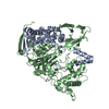
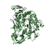
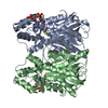
 Archaeoglobus fulgidus (archaea)
 X-RAY DIFFRACTION /
  Authors
 Citation
 Structure visualization
 Downloads & links
 Other downloads
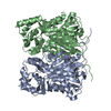

 PDBj
PDBj Assembly


 Mass: 94.971 Da / Num. of mol.: 2 / Source method: obtained synthetically / Formula: PO4
 Mass: 106.120 Da / Num. of mol.: 1 / Source method: obtained synthetically / Formula: C4H10O3
 Mass: 18.015 Da / Num. of mol.: 76 / Source method: isolated from a natural source / Formula: H2O
 Sample preparation
 / Beamline: BL11-1
 Processing