 Movie
Movie Controller


+ Open data

- Basic information
| Entry | Database: PDB / ID: 7crv | ||||||
|---|---|---|---|---|---|---|---|
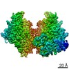
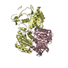
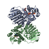
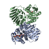
| Title | Crystal structure of rNLRP1-FIIND | ||||||
 Components Components | (NLR family protein 1) x 2 | ||||||
 Keywords Keywords | IMMUNE SYSTEM | ||||||
| Function / homology |  Function and homology information Function and homology informationnegative regulation of cellular defense response / canonical inflammasome complex / positive regulation of pyroptotic inflammatory response / programmed cell death / Hydrolases; Acting on peptide bonds (peptidases) / peptidase activity / scaffold protein binding / regulation of apoptotic process / inflammatory response / innate immune response ...negative regulation of cellular defense response / canonical inflammasome complex / positive regulation of pyroptotic inflammatory response / programmed cell death / Hydrolases; Acting on peptide bonds (peptidases) / peptidase activity / scaffold protein binding / regulation of apoptotic process / inflammatory response / innate immune response / neuronal cell body / proteolysis / ATP binding / nucleus Similarity search - Function | ||||||
| Biological species |  | ||||||
| Method |  X-RAY DIFFRACTION / SYNCHROTRON / MOLECULAR REPLACEMENT / Resolution: 2 Å X-RAY DIFFRACTION / SYNCHROTRON / MOLECULAR REPLACEMENT / Resolution: 2 Å | ||||||
 Authors Authors | Huang, M.H. / Zhang, X.X. / Wang, J. / Chai, J.J. | ||||||
| Funding support |  China, 1items China, 1items
| ||||||
 Citation Citation | Journal: Nature / Year: 2021 Title: Structural and biochemical mechanisms of NLRP1 inhibition by DPP9. Authors: Menghang Huang / Xiaoxiao Zhang / Gee Ann Toh / Qin Gong / Jia Wang / Zhifu Han / Bin Wu / Franklin Zhong / Jijie Chai /   Abstract: Nucleotide-binding domain, leucine-rich repeat receptors (NLRs) mediate innate immunity by forming inflammasomes. Activation of the NLR protein NLRP1 requires autocleavage within its function-to-find ...Nucleotide-binding domain, leucine-rich repeat receptors (NLRs) mediate innate immunity by forming inflammasomes. Activation of the NLR protein NLRP1 requires autocleavage within its function-to-find domain (FIIND). In resting cells, the dipeptidyl peptidases DPP8 and DPP9 interact with the FIIND of NLRP1 and suppress spontaneous NLRP1 activation; however, the mechanisms through which this occurs remain unknown. Here we present structural and biochemical evidence that full-length rat NLRP1 (rNLRP1) and rat DPP9 (rDPP9) form a 2:1 complex that contains an autoinhibited rNLRP1 molecule and an active UPA-CARD fragment of rNLRP1. The ZU5 domain is required not only for autoinhibition of rNLRP1 but also for assembly of the 2:1 complex. Formation of the complex prevents UPA-mediated higher-order oligomerization of UPA-CARD fragments and strengthens ZU5-mediated NLRP1 autoinhibition. Structure-guided biochemical and functional assays show that both NLRP1 binding and enzymatic activity are required for DPP9 to suppress NLRP1 in human cells. Together, our data reveal the mechanism of DPP9-mediated inhibition of NLRP1 and shed light on the activation of the NLRP1 inflammasome. | ||||||
| History |
|
- Structure visualization
| Structure viewer | Molecule: MolmilJmol/JSmol |
|---|
- Downloads & links
-Download
| PDBx/mmCIF format | 7crv.cif.gz | 152.8 KB | Display | PDBx/mmCIF format |
|---|---|---|---|---|
| PDB format | pdb7crv.ent.gz | 96.3 KB | Display | PDB format |
| PDBx/mmJSON format | 7crv.json.gz | Tree view | PDBx/mmJSON format | |
| Others |  Other downloads Other downloads |
-Validation report
| Arichive directory | https://data.pdbj.org/pub/pdb/validation_reports/cr/7crvftp://data.pdbj.org/pub/pdb/validation_reports/cr/7crv | HTTPS FTP |
|---|
-Related structure data
| Related structure data |  7crwC  3g5bS S: Starting model for refinement C: citing same article ( |
|---|---|
| Similar structure data |


-Links
 PDBj
PDBj


- Assembly
| Deposited unit | 
| ||||||||||||
|---|---|---|---|---|---|---|---|---|---|---|---|---|---|
| 1 |
| ||||||||||||
| Unit cell |
|
-Components
| #1: Protein | Mass: 20764.316 Da / Num. of mol.: 2 / Fragment: ZU5 domain Source method: isolated from a genetically manipulated source Source: (gene. exp.)   Spodoptera frugiperda (fall armyworm) / References: UniProt: D9I2G3 Spodoptera frugiperda (fall armyworm) / References: UniProt: D9I2G3#2: Protein | Mass: 17649.637 Da / Num. of mol.: 2 / Fragment: UPA domain Source method: isolated from a genetically manipulated source Source: (gene. exp.) Spodoptera frugiperda (fall armyworm) / References: UniProt: D9I2G3 |
|---|
-Experimental details
-Experiment
| Experiment | Method: X-RAY DIFFRACTION / Number of used crystals: 1 |
|---|
- Sample preparation
| Crystal | Density Matthews: 2.07 Å3/Da / Density % sol: 40.72 % |
|---|---|
| Crystal grow | Temperature: 291 K / Method: vapor diffusion, hanging drop Details: 1.0M Ammonium sulfate, 0.1M Bis-Tris pH 5.5, 1% w/v polyethylene glycol 3350 |
-Data collection
| Diffraction | Mean temperature: 100 K / Serial crystal experiment: N |
|---|---|
| Diffraction source | Source: SYNCHROTRON / Site: SSRF / Beamline: BL17U1 / Wavelength: 0.979 Å |
| Detector | Type: DECTRIS EIGER X 16M / Detector: PIXEL / Date: Oct 25, 2019 |
| Radiation | Protocol: SINGLE WAVELENGTH / Monochromatic (M) / Laue (L): M / Scattering type: x-ray |
| Radiation wavelength | Wavelength: 0.979 Å / Relative weight: 1 |
| Reflection | Resolution: 2→50 Å / Num. obs: 43870 / % possible obs: 99.9 % / Redundancy: 19.3 % / Biso Wilson estimate: 31.87 Å2 / CC1/2: 0.997 / Net I/σ(I): 30.9 |
| Reflection shell | Resolution: 2→2.03 Å / Num. unique obs: 4107 / CC1/2: 0.776 |
- Processing
| Software |
| |||||||||||||||||||||||||||||||||||||||||||||||||||||||||||||||||||||||||||||||||||||||||||||||||||||||||||||||||||||||
|---|---|---|---|---|---|---|---|---|---|---|---|---|---|---|---|---|---|---|---|---|---|---|---|---|---|---|---|---|---|---|---|---|---|---|---|---|---|---|---|---|---|---|---|---|---|---|---|---|---|---|---|---|---|---|---|---|---|---|---|---|---|---|---|---|---|---|---|---|---|---|---|---|---|---|---|---|---|---|---|---|---|---|---|---|---|---|---|---|---|---|---|---|---|---|---|---|---|---|---|---|---|---|---|---|---|---|---|---|---|---|---|---|---|---|---|---|---|---|---|---|
| Refinement | Method to determine structure: MOLECULAR REPLACEMENT Starting model: 3G5B Resolution: 2→28.76 Å / SU ML: 0.2651 / Cross valid method: FREE R-VALUE / σ(F): 1.36 / Phase error: 28.6481 Stereochemistry target values: GeoStd + Monomer Library + CDL v1.2
| |||||||||||||||||||||||||||||||||||||||||||||||||||||||||||||||||||||||||||||||||||||||||||||||||||||||||||||||||||||||
| Solvent computation | Shrinkage radii: 0.9 Å / VDW probe radii: 1.11 Å / Solvent model: FLAT BULK SOLVENT MODEL | |||||||||||||||||||||||||||||||||||||||||||||||||||||||||||||||||||||||||||||||||||||||||||||||||||||||||||||||||||||||
| Displacement parameters | Biso mean: 37.34 Å2 | |||||||||||||||||||||||||||||||||||||||||||||||||||||||||||||||||||||||||||||||||||||||||||||||||||||||||||||||||||||||
| Refinement step | Cycle: LAST / Resolution: 2→28.76 Å
| |||||||||||||||||||||||||||||||||||||||||||||||||||||||||||||||||||||||||||||||||||||||||||||||||||||||||||||||||||||||
| Refine LS restraints |
| |||||||||||||||||||||||||||||||||||||||||||||||||||||||||||||||||||||||||||||||||||||||||||||||||||||||||||||||||||||||
| LS refinement shell |
|
