| Entry | Database: PDB / ID: 4beg
|
|---|
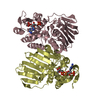
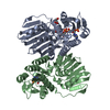
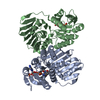
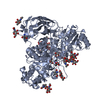
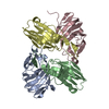
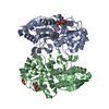
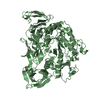
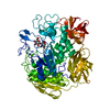
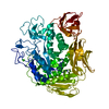
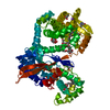
| Title | Structure of Rv2140c, a phosphatidyl-ethanolamine binding protein from Mycobacterium tuberculosis in complex with sulphate |
|---|
 Components Components | PHOSPHATIDYLETHANOLAMINE BINDING PROTEIN |
|---|
 Keywords Keywords | LIPID BINDING PROTEIN / LIPID-BINDING PROTEIN / PEBP / RKIP |
|---|
| Function / homology |  Function and homology information Function and homology information
YbhB/YbcL / Phosphatidylethanolamine-binding Protein / PEBP-like / Phosphatidylethanolamine-binding protein / Phosphatidylethanolamine-binding protein / PEBP-like superfamily / Alpha-Beta Complex / Alpha BetaSimilarity search - Domain/homology |
|---|
| Biological species |   MYCOBACTERIUM TUBERCULOSIS (bacteria) MYCOBACTERIUM TUBERCULOSIS (bacteria) |
|---|
| Method |  X-RAY DIFFRACTION / SYNCHROTRON / MOLECULAR REPLACEMENT / Resolution: 1.42 Å X-RAY DIFFRACTION / SYNCHROTRON / MOLECULAR REPLACEMENT / Resolution: 1.42 Å |
|---|
 Authors Authors | Holton, S.J. / Williams, M. |
|---|
 Citation Citation | Journal: FEBS Lett. / Year: 2013
Title: Structural and Biochemical Characterization of Rv2140C, a Phosphatidylethanolamine-Binding Protein from Mycobacterium Tuberculosis.
Authors: Eulenburg, G. / Higman, V.A. / Diehl, A. / Wilmanns, M. / Holton, S.J. |
|---|
| History | | Deposition | Mar 9, 2013 | Deposition site: PDBE / Processing site: PDBE |
|---|
| Revision 1.0 | Aug 14, 2013 | Provider: repository / Type: Initial release |
|---|
| Revision 1.1 | Sep 25, 2013 | Group: Database references |
|---|
| Revision 1.2 | May 22, 2019 | Group: Data collection / Other / Refinement description
Category: pdbx_database_proc / pdbx_database_status / refine
Item: _pdbx_database_status.recvd_author_approval / _refine.pdbx_ls_cross_valid_method |
|---|
| Revision 1.3 | Jul 17, 2019 | Group: Data collection / Category: diffrn_source / Item: _diffrn_source.pdbx_synchrotron_site |
|---|
| Revision 1.4 | Dec 20, 2023 | Group: Data collection / Database references ...Data collection / Database references / Derived calculations / Other / Refinement description
Category: chem_comp_atom / chem_comp_bond ...chem_comp_atom / chem_comp_bond / database_2 / pdbx_database_status / pdbx_initial_refinement_model / struct_site
Item: _database_2.pdbx_DOI / _database_2.pdbx_database_accession ..._database_2.pdbx_DOI / _database_2.pdbx_database_accession / _pdbx_database_status.status_code_sf / _struct_site.pdbx_auth_asym_id / _struct_site.pdbx_auth_comp_id / _struct_site.pdbx_auth_seq_id |
|---|
|
|---|
 Movie
Movie Controller

 Yorodumi
 Open data
 Basic information
 Structure visualization
 Downloads & links
 Other downloads
 PDBj
PDBj Assembly
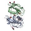


 Mass: 96.063 Da / Num. of mol.: 2 / Source method: obtained synthetically / Formula: SO4
 Mass: 92.094 Da / Num. of mol.: 6 / Source method: obtained synthetically / Formula: C3H8O3
 Mass: 18.015 Da / Num. of mol.: 345 / Source method: isolated from a natural source / Formula: H2O
 Sample preparation
 / Beamline: BW7B / Wavelength: 0.84
  Processing