 Movie
Movie Controller


+ Open data

- Basic information
| Entry | Database: PDB / ID: 5tdx | ||||||
|---|---|---|---|---|---|---|---|
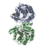
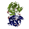
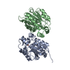
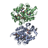
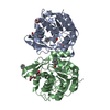
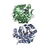
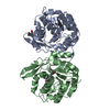
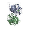
| Title | Resurrected Ancestral Hydroxynitrile Lyase from Flowering Plants | ||||||
 Components Components | Ancestral Hydroxynitrile Lyase 1 | ||||||
 Keywords Keywords | LYASE / HNL Ancestral Hydroxynitrile lyase | ||||||
| Function / homology | Alpha/Beta hydrolase fold, catalytic domain / Rossmann fold / 3-Layer(aba) Sandwich / Alpha Beta Function and homology information Function and homology information | ||||||
| Biological species | synthetic construct (others) | ||||||
| Method |  X-RAY DIFFRACTION / SYNCHROTRON / MOLECULAR REPLACEMENT / molecular replacement / Resolution: 1.96 Å X-RAY DIFFRACTION / SYNCHROTRON / MOLECULAR REPLACEMENT / molecular replacement / Resolution: 1.96 Å | ||||||
 Authors Authors | Jones, B.J. / Evans, R. / Wilmot, C.M. / Kazlauskas, R.J. | ||||||
| Funding support |  United States, 1items United States, 1items
| ||||||
 Citation Citation | Journal: Plos One / Year: 2020 Title: Larger active site in an ancestral hydroxynitrile lyase increases catalytically promiscuous esterase activity. Authors: Jones, B.J. / Evans 3rd, R.L. / Mylrea, N.J. / Chaudhury, D. / Luo, C. / Guan, B. / Pierce, C.T. / Gordon, W.R. / Wilmot, C.M. / Kazlauskas, R.J. | ||||||
| History |
|
- Structure visualization
| Structure viewer | Molecule: MolmilJmol/JSmol |
|---|
- Downloads & links
-Download
| PDBx/mmCIF format | 5tdx.cif.gz | 427.9 KB | Display | PDBx/mmCIF format |
|---|---|---|---|---|
| PDB format | pdb5tdx.ent.gz | 354.1 KB | Display | PDB format |
| PDBx/mmJSON format | 5tdx.json.gz | Tree view | PDBx/mmJSON format | |
| Others |  Other downloads Other downloads |
-Validation report
| Arichive directory | https://data.pdbj.org/pub/pdb/validation_reports/td/5tdxftp://data.pdbj.org/pub/pdb/validation_reports/td/5tdx | HTTPS FTP |
|---|
-Related structure data
| Related structure data |  1yb6S S: Starting model for refinement |
|---|---|
| Similar structure data |


-Links
 PDBj
PDBj- Assembly
| Deposited unit | 
| ||||||||
|---|---|---|---|---|---|---|---|---|---|
| 1 | 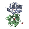
| ||||||||
| 2 | 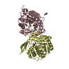
| ||||||||
| Unit cell |
|
-Components
| #1: Protein | Mass: 31119.217 Da / Num. of mol.: 4 Source method: isolated from a genetically manipulated source Source: (gene. exp.) synthetic construct (others) / Plasmid: pET21a / Production host:  #2: Chemical | ChemComp-GOL /   Mass: 92.094 Da / Num. of mol.: 6 / Source method: obtained synthetically / Formula: C3H8O3 Mass: 92.094 Da / Num. of mol.: 6 / Source method: obtained synthetically / Formula: C3H8O3#3: Water | ChemComp-HOH / |  Mass: 18.015 Da / Num. of mol.: 192 / Source method: isolated from a natural source / Formula: H2O Mass: 18.015 Da / Num. of mol.: 192 / Source method: isolated from a natural source / Formula: H2O |
|---|
-Experimental details
-Experiment
| Experiment | Method: X-RAY DIFFRACTION / Number of used crystals: 1 |
|---|
- Sample preparation
| Crystal | Density Matthews: 2.44 Å3/Da / Density % sol: 49.66 % / Description: Octohedral, 220 um point to point |
|---|---|
| Crystal grow | Temperature: 293 K / Method: vapor diffusion, hanging drop / pH: 6.8 Details: 1:1 mix of protein (in 5mM BES buffer) with mother liquor (1.1 Molar Ammonium Citrate, 50 mM bis-tris propane, pH 6.8) Crystals formed after 2-3 weeks at 20 degrees C. Crystals were cryo- ...Details: 1:1 mix of protein (in 5mM BES buffer) with mother liquor (1.1 Molar Ammonium Citrate, 50 mM bis-tris propane, pH 6.8) Crystals formed after 2-3 weeks at 20 degrees C. Crystals were cryo-protected in 5% glycerol in mother liquor for 10 seconds then flash frozen in LN2. |
-Data collection
| Diffraction | Mean temperature: 100 K |
|---|---|
| Diffraction source | Source: SYNCHROTRON / Site: APS / Beamline: 23-ID-B / Wavelength: 1.0332 Å |
| Detector | Type: MAR scanner 300 mm plate / Detector: IMAGE PLATE / Date: Oct 30, 2014 |
| Radiation | Monochromator: Si(111) / Protocol: SINGLE WAVELENGTH / Monochromatic (M) / Laue (L): M / Scattering type: x-ray |
| Radiation wavelength | Wavelength: 1.0332 Å / Relative weight: 1 |
| Reflection | Resolution: 1.96→29.32 Å / Num. obs: 86348 / % possible obs: 99.3 % / Redundancy: 8.5 % / CC1/2: 0.998 / Rmerge(I) obs: 0.097 / Net I/σ(I): 13.8 |
| Reflection shell | Resolution: 1.96→1.99 Å / Redundancy: 5.8 % / Rmerge(I) obs: 0.68 / CC1/2: 0.772 / % possible all: 88.3 |
-Phasing
| Phasing | Method: molecular replacement | |||||||||
|---|---|---|---|---|---|---|---|---|---|---|
| Phasing MR | Model details: Phaser MODE: MR_AUTO
|
- Processing
| Software |
| |||||||||||||||||||||||||||||||||||||||||||||||||||||||||||||||||||||||||||||||||||||||||||||||||||||||||||||||||||||||||||||
|---|---|---|---|---|---|---|---|---|---|---|---|---|---|---|---|---|---|---|---|---|---|---|---|---|---|---|---|---|---|---|---|---|---|---|---|---|---|---|---|---|---|---|---|---|---|---|---|---|---|---|---|---|---|---|---|---|---|---|---|---|---|---|---|---|---|---|---|---|---|---|---|---|---|---|---|---|---|---|---|---|---|---|---|---|---|---|---|---|---|---|---|---|---|---|---|---|---|---|---|---|---|---|---|---|---|---|---|---|---|---|---|---|---|---|---|---|---|---|---|---|---|---|---|---|---|---|
| Refinement | Method to determine structure: MOLECULAR REPLACEMENT Starting model: 1YB6 Resolution: 1.96→29.32 Å / Cor.coef. Fo:Fc: 0.966 / Cor.coef. Fo:Fc free: 0.945 / SU B: 8.339 / SU ML: 0.113 / Cross valid method: THROUGHOUT / σ(F): 0 / ESU R: 0.152 / ESU R Free: 0.148 / Details: HYDROGENS HAVE BEEN ADDED IN THE RIDING POSITIONS
| |||||||||||||||||||||||||||||||||||||||||||||||||||||||||||||||||||||||||||||||||||||||||||||||||||||||||||||||||||||||||||||
| Solvent computation | Ion probe radii: 0.8 Å / Shrinkage radii: 0.8 Å / VDW probe radii: 1.2 Å | |||||||||||||||||||||||||||||||||||||||||||||||||||||||||||||||||||||||||||||||||||||||||||||||||||||||||||||||||||||||||||||
| Displacement parameters | Biso max: 112.94 Å2 / Biso mean: 38.133 Å2 / Biso min: 15.28 Å2
| |||||||||||||||||||||||||||||||||||||||||||||||||||||||||||||||||||||||||||||||||||||||||||||||||||||||||||||||||||||||||||||
| Refinement step | Cycle: final / Resolution: 1.96→29.32 Å
| |||||||||||||||||||||||||||||||||||||||||||||||||||||||||||||||||||||||||||||||||||||||||||||||||||||||||||||||||||||||||||||
| Refine LS restraints |
| |||||||||||||||||||||||||||||||||||||||||||||||||||||||||||||||||||||||||||||||||||||||||||||||||||||||||||||||||||||||||||||
| LS refinement shell | Resolution: 1.956→2.007 Å / Total num. of bins used: 20
| |||||||||||||||||||||||||||||||||||||||||||||||||||||||||||||||||||||||||||||||||||||||||||||||||||||||||||||||||||||||||||||
| Refinement TLS params. | Method: refined / Refine-ID: X-RAY DIFFRACTION
| |||||||||||||||||||||||||||||||||||||||||||||||||||||||||||||||||||||||||||||||||||||||||||||||||||||||||||||||||||||||||||||
| Refinement TLS group |
|
