 Movie
Movie Controller

[English] 日本語
 Yorodumi
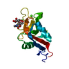
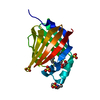
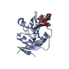
- PDB-6pws: Crystal structure of the cow C-type carbohydrate-recognition doma... -
+ Open data

- Basic information
| Entry | Database: PDB / ID: 6pws | ||||||
|---|---|---|---|---|---|---|---|
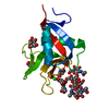
| Title | Crystal structure of the cow C-type carbohydrate-recognition domain of CD23 in the presence of alpha-methyl mannoside | ||||||
 Components Components | Fc fragment of IgE receptor II | ||||||
 Keywords Keywords | SUGAR BINDING PROTEIN / CRD / Receptor / Lectin / Metal-Binding | ||||||
| Function / homology |  Function and homology information Function and homology informationpattern recognition receptor activity / carbohydrate binding / immune response / external side of plasma membrane / metal ion binding Similarity search - Function | ||||||
| Biological species |  | ||||||
| Method |  X-RAY DIFFRACTION / SYNCHROTRON / MOLECULAR REPLACEMENT / molecular replacement / Resolution: 1 Å X-RAY DIFFRACTION / SYNCHROTRON / MOLECULAR REPLACEMENT / molecular replacement / Resolution: 1 Å | ||||||
 Authors Authors | Weis, W.I. / Feinberg, H. | ||||||
| Funding support |  United Kingdom, 1items United Kingdom, 1items
| ||||||
 Citation Citation | Journal: J.Biol.Chem. / Year: 2019 Title: CD23 is a glycan-binding receptor in some mammalian species. Authors: Jegouzo, S.A.F. / Feinberg, H. / Morrison, A.G. / Holder, A. / May, A. / Huang, Z. / Jiang, L. / Lasanajak, Y. / Smith, D.F. / Werling, D. / Drickamer, K. / Weis, W.I. / Taylor, M.E. | ||||||
| History |
|
- Structure visualization
| Structure viewer | Molecule: MolmilJmol/JSmol |
|---|
- Downloads & links
-Download
| PDBx/mmCIF format | 6pws.cif.gz | 97 KB | Display | PDBx/mmCIF format |
|---|---|---|---|---|
| PDB format | pdb6pws.ent.gz | 72 KB | Display | PDB format |
| PDBx/mmJSON format | 6pws.json.gz | Tree view | PDBx/mmJSON format | |
| Others |  Other downloads Other downloads |
-Validation report
| Arichive directory | https://data.pdbj.org/pub/pdb/validation_reports/pw/6pwsftp://data.pdbj.org/pub/pdb/validation_reports/pw/6pws | HTTPS FTP |
|---|
-Related structure data
| Related structure data |  6pwrC  6pwtC  2h2tS S: Starting model for refinement C: citing same article ( |
|---|---|
| Similar structure data |


-Links
 PDBj
PDBj


- Assembly
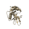
| Deposited unit | 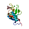
| ||||||||
|---|---|---|---|---|---|---|---|---|---|
| 1 |
| ||||||||
| Unit cell |
|
-Components
| #1: Protein | Mass: 15349.125 Da / Num. of mol.: 1 Source method: isolated from a genetically manipulated source Source: (gene. exp.)  | ||||||||||
|---|---|---|---|---|---|---|---|---|---|---|---|
| #2: Chemical |   Mass: 40.078 Da / Num. of mol.: 2 / Source method: obtained synthetically / Formula: Ca Mass: 40.078 Da / Num. of mol.: 2 / Source method: obtained synthetically / Formula: Ca#3: Sugar | ChemComp-MMA / | 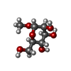  Type: D-saccharide / Mass: 194.182 Da / Num. of mol.: 1 Type: D-saccharide / Mass: 194.182 Da / Num. of mol.: 1Source method: isolated from a genetically manipulated source Formula: C7H14O6 #4: Water | ChemComp-HOH / |  Mass: 18.015 Da / Num. of mol.: 244 / Source method: isolated from a natural source / Formula: H2O Mass: 18.015 Da / Num. of mol.: 244 / Source method: isolated from a natural source / Formula: H2OHas ligand of interest | N | Has protein modification | Y | Sequence details | Residue numbering based on NCBI Reference Sequence: XP_002688905.2 | |
-Experimental details
-Experiment
| Experiment | Method: X-RAY DIFFRACTION / Number of used crystals: 1 |
|---|
- Sample preparation
| Crystal | Density Matthews: 2.05 Å3/Da / Density % sol: 40.13 % |
|---|---|
| Crystal grow | Temperature: 290 K / Method: vapor diffusion, hanging drop / pH: 5.5 Details: protein solution: 4.2 mg/ml protein, 5 mM CaCl2, 10 mM Tris-Cl, pH 8.0, 25 mM NaCl, and 50 mM alpha-methyl mannoside. reservoir solution: 16% polyethylene glycol 4K, 0.1 MES, pH 5.5. |
-Data collection
| Diffraction | Mean temperature: 100 K / Serial crystal experiment: N | ||||||||||||||||||||||||||||||
|---|---|---|---|---|---|---|---|---|---|---|---|---|---|---|---|---|---|---|---|---|---|---|---|---|---|---|---|---|---|---|---|
| Diffraction source | Source: SYNCHROTRON / Site: SSRL  / Beamline: BL12-2 / Wavelength: 0.8157 Å / Beamline: BL12-2 / Wavelength: 0.8157 Å | ||||||||||||||||||||||||||||||
| Detector | Type: DECTRIS PILATUS 6M / Detector: PIXEL / Date: Dec 16, 2018 | ||||||||||||||||||||||||||||||
| Radiation | Protocol: SINGLE WAVELENGTH / Monochromatic (M) / Laue (L): M / Scattering type: x-ray | ||||||||||||||||||||||||||||||
| Radiation wavelength | Wavelength: 0.8157 Å / Relative weight: 1 | ||||||||||||||||||||||||||||||
| Reflection | Resolution: 1→38.55 Å / Num. obs: 67049 / % possible obs: 99.7 % / Redundancy: 6.4 % / CC1/2: 0.998 / Rmerge(I) obs: 0.033 / Rpim(I) all: 0.014 / Rrim(I) all: 0.036 / Net I/σ(I): 33.5 / Num. measured all: 429513 / Scaling rejects: 244 | ||||||||||||||||||||||||||||||
| Reflection shell | Diffraction-ID: 1
|
-Phasing
| Phasing | Method: molecular replacement |
|---|
- Processing
| Software |
| ||||||||||||||||||||||||||||||||||||||||||||||||||||||||||||||||||||||||||||||||||||||||||||||||||||||||||||||||||||||||||||||||||||||||||||||||||||||
|---|---|---|---|---|---|---|---|---|---|---|---|---|---|---|---|---|---|---|---|---|---|---|---|---|---|---|---|---|---|---|---|---|---|---|---|---|---|---|---|---|---|---|---|---|---|---|---|---|---|---|---|---|---|---|---|---|---|---|---|---|---|---|---|---|---|---|---|---|---|---|---|---|---|---|---|---|---|---|---|---|---|---|---|---|---|---|---|---|---|---|---|---|---|---|---|---|---|---|---|---|---|---|---|---|---|---|---|---|---|---|---|---|---|---|---|---|---|---|---|---|---|---|---|---|---|---|---|---|---|---|---|---|---|---|---|---|---|---|---|---|---|---|---|---|---|---|---|---|---|---|---|
| Refinement | Method to determine structure: MOLECULAR REPLACEMENT Starting model: 2H2T Resolution: 1→29.525 Å / Cross valid method: THROUGHOUT
| ||||||||||||||||||||||||||||||||||||||||||||||||||||||||||||||||||||||||||||||||||||||||||||||||||||||||||||||||||||||||||||||||||||||||||||||||||||||
| Displacement parameters | Biso max: 52.11 Å2 / Biso mean: 10.2 Å2 / Biso min: 3.61 Å2 | ||||||||||||||||||||||||||||||||||||||||||||||||||||||||||||||||||||||||||||||||||||||||||||||||||||||||||||||||||||||||||||||||||||||||||||||||||||||
| Refinement step | Cycle: final / Resolution: 1→29.525 Å
| ||||||||||||||||||||||||||||||||||||||||||||||||||||||||||||||||||||||||||||||||||||||||||||||||||||||||||||||||||||||||||||||||||||||||||||||||||||||
| LS refinement shell | Refine-ID: X-RAY DIFFRACTION / Rfactor Rfree error: 0
|
