[English] 日本語
Yorodumi
- PDB-2yhf: 1.9 Angstrom Crystal Structure of CLEC5A -

+
Open data


ID or keywords:

Loading...

-
Basic information

Entry
Database: PDB / ID: 2yhf
Title1.9 Angstrom Crystal Structure of CLEC5A
ComponentsC-TYPE LECTIN DOMAIN FAMILY 5 MEMBER A
KeywordsIMMUNE SYSTEM
Function / homology
Function and homology information


positive regulation of cytokine production => GO:0001819 / neutrophil degranulation / negative regulation of myeloid cell apoptotic process / myeloid cell differentiation / osteoblast development / plasma membrane => GO:0005886 / tertiary granule membrane / cellular defense response / specific granule membrane / DAP12 interactions ...positive regulation of cytokine production => GO:0001819 / neutrophil degranulation / negative regulation of myeloid cell apoptotic process / myeloid cell differentiation / osteoblast development / plasma membrane => GO:0005886 / tertiary granule membrane / cellular defense response / specific granule membrane / DAP12 interactions / virus receptor activity / carbohydrate binding / innate immune response / Neutrophil degranulation / negative regulation of apoptotic process / cell surface / signal transduction / plasma membrane / cytosol
Similarity search - Function
Natural killer cell receptor-like, C-type lectin-like domain / Mannose-Binding Protein A; Chain A / Mannose-Binding Protein A, subunit A / Lectin C-type domain / C-type lectin domain profile. / C-type lectin-like / C-type lectin (CTL) or carbohydrate-recognition domain (CRD) / C-type lectin-like/link domain superfamily / C-type lectin fold / Roll / Alpha Beta
Similarity search - Domain/homology
C-type lectin domain family 5 member A
Similarity search - Component
Biological speciesHOMO SAPIENS (human)
MethodX-RAY DIFFRACTION / SYNCHROTRON / MOLECULAR REPLACEMENT / Resolution: 1.9 Å
AuthorsWatson, A.A. / Lebedev, A.A. / Murshudov, G.M. / Vagin, A.A. / Hall, B.A. / O'Callaghan, C.A.
CitationJournal: J.Biol.Chem. / Year: 2011
Title: Structural Flexibility of the Macrophage Dengue Virus Receptor Clec5A: Implications for Ligand Binding and Signaling.
Authors: Watson, A.A. / Lebedev, A.A. / Hall, B.A. / Fenton-May, A.E. / Vagin, A.A. / Dejnirattisai, W. / Felce, J. / Mongkolsapaya, J. / Palma, A.S. / Liu, Y. / Feizi, T. / Screaton, G.R. / ...Authors: Watson, A.A. / Lebedev, A.A. / Hall, B.A. / Fenton-May, A.E. / Vagin, A.A. / Dejnirattisai, W. / Felce, J. / Mongkolsapaya, J. / Palma, A.S. / Liu, Y. / Feizi, T. / Screaton, G.R. / Murshudov, G.N. / O'Callaghan, C.A.
History
DepositionApr 30, 2011Deposition site: PDBE / Processing site: PDBE
Revision 1.0May 11, 2011Provider: repository / Type: Initial release
Revision 1.1Jul 7, 2011Group: Version format compliance
Revision 1.2Jul 13, 2011Group: Version format compliance
Revision 1.3Dec 20, 2023Group: Data collection / Database references ...Data collection / Database references / Other / Refinement description
Category: chem_comp_atom / chem_comp_bond ...chem_comp_atom / chem_comp_bond / database_2 / pdbx_database_status / pdbx_initial_refinement_model
Item: _database_2.pdbx_DOI / _database_2.pdbx_database_accession / _pdbx_database_status.status_code_sf

-
Structure visualization

Structure viewerMolecule:
MolmilJmol/JSmol

Downloads & links

-
Assembly

Deposited unit
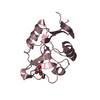
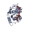
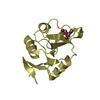
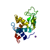
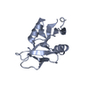
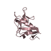
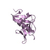
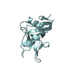
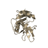
A: C-TYPE LECTIN DOMAIN FAMILY 5 MEMBER A
B: C-TYPE LECTIN DOMAIN FAMILY 5 MEMBER A
C: C-TYPE LECTIN DOMAIN FAMILY 5 MEMBER A
D: C-TYPE LECTIN DOMAIN FAMILY 5 MEMBER A
E: C-TYPE LECTIN DOMAIN FAMILY 5 MEMBER A
F: C-TYPE LECTIN DOMAIN FAMILY 5 MEMBER A
G: C-TYPE LECTIN DOMAIN FAMILY 5 MEMBER A
H: C-TYPE LECTIN DOMAIN FAMILY 5 MEMBER A
I: C-TYPE LECTIN DOMAIN FAMILY 5 MEMBER A


Theoretical massNumber of molelcules
Total (without water)124,5029
Polymers124,5029
Non-polymers00
Water16,934940
1
A: C-TYPE LECTIN DOMAIN FAMILY 5 MEMBER A


Theoretical massNumber of molelcules
Total (without water)13,8341
Polymers13,8341
Non-polymers00
Water181
TypeNameSymmetry operationNumber
identity operation1_555x,y,z1
MethodPISA
2
B: C-TYPE LECTIN DOMAIN FAMILY 5 MEMBER A


Theoretical massNumber of molelcules
Total (without water)13,8341
Polymers13,8341
Non-polymers00
Water181
TypeNameSymmetry operationNumber
identity operation1_555x,y,z1
MethodPISA
3
C: C-TYPE LECTIN DOMAIN FAMILY 5 MEMBER A


Theoretical massNumber of molelcules
Total (without water)13,8341
Polymers13,8341
Non-polymers00
Water181
TypeNameSymmetry operationNumber
identity operation1_555x,y,z1
MethodPISA
4
D: C-TYPE LECTIN DOMAIN FAMILY 5 MEMBER A


Theoretical massNumber of molelcules
Total (without water)13,8341
Polymers13,8341
Non-polymers00
Water181
TypeNameSymmetry operationNumber
identity operation1_555x,y,z1
MethodPISA
5
E: C-TYPE LECTIN DOMAIN FAMILY 5 MEMBER A


Theoretical massNumber of molelcules
Total (without water)13,8341
Polymers13,8341
Non-polymers00
Water181
TypeNameSymmetry operationNumber
identity operation1_555x,y,z1
MethodPISA
6
F: C-TYPE LECTIN DOMAIN FAMILY 5 MEMBER A


Theoretical massNumber of molelcules
Total (without water)13,8341
Polymers13,8341
Non-polymers00
Water181
TypeNameSymmetry operationNumber
identity operation1_555x,y,z1
MethodPISA
7
G: C-TYPE LECTIN DOMAIN FAMILY 5 MEMBER A


Theoretical massNumber of molelcules
Total (without water)13,8341
Polymers13,8341
Non-polymers00
Water181
TypeNameSymmetry operationNumber
identity operation1_555x,y,z1
MethodPISA
8
H: C-TYPE LECTIN DOMAIN FAMILY 5 MEMBER A


Theoretical massNumber of molelcules
Total (without water)13,8341
Polymers13,8341
Non-polymers00
Water181
TypeNameSymmetry operationNumber
identity operation1_555x,y,z1
MethodPISA
9
I: C-TYPE LECTIN DOMAIN FAMILY 5 MEMBER A


Theoretical massNumber of molelcules
Total (without water)13,8341
Polymers13,8341
Non-polymers00
Water181
TypeNameSymmetry operationNumber
identity operation1_555x,y,z1
MethodPISA
Unit cell
Length a, b, c (Å)109.109, 109.109, 84.879
Angle α, β, γ (deg.)90.00, 90.00, 120.00
Int Tables number144
Space group name H-MP31

-
Components

#1: Protein
C-TYPE LECTIN DOMAIN FAMILY 5 MEMBER A / C-TYPE LECTIN SUPERFAMILY MEMBER 5 / MYELOID DAP12-ASSOCIATING LECTIN 1 / MDL-1 / CL3


Mass: 13833.537 Da / Num. of mol.: 9 / Fragment: RESIDUES 70-187 / Mutation: YES
Source method: isolated from a genetically manipulated source
Source: (gene. exp.) HOMO SAPIENS (human) / Cell: PERIPHERAL BLOOD MONONUCLEAR CELLS / Production host: ESCHERICHIA COLI (E. coli) / Strain (production host): BL21(DE3) / Variant (production host): PLYSS / References: UniProt: Q9NY25
#2: Water ChemComp-HOH / water


Mass: 18.015 Da / Num. of mol.: 940 / Source method: isolated from a natural source / Formula: H2O
Compound detailsENGINEERED RESIDUE IN CHAIN A, VAL 70 TO MET ENGINEERED RESIDUE IN CHAIN B, VAL 70 TO MET ...ENGINEERED RESIDUE IN CHAIN A, VAL 70 TO MET ENGINEERED RESIDUE IN CHAIN B, VAL 70 TO MET ENGINEERED RESIDUE IN CHAIN C, VAL 70 TO MET ENGINEERED RESIDUE IN CHAIN D, VAL 70 TO MET ENGINEERED RESIDUE IN CHAIN E, VAL 70 TO MET ENGINEERED RESIDUE IN CHAIN F, VAL 70 TO MET ENGINEERED RESIDUE IN CHAIN G, VAL 70 TO MET ENGINEERED RESIDUE IN CHAIN H, VAL 70 TO MET ENGINEERED RESIDUE IN CHAIN I, VAL 70 TO MET

-
Experimental details

-
Experiment

ExperimentMethod: X-RAY DIFFRACTION / Number of used crystals: 1

-
Sample preparation

CrystalDensity Matthews: 2.35 Å3/Da / Density % sol: 47.56 % / Description: NONE
Crystal growpH: 8 / Details: 100MM TRIS PH8.0, 30% PEG 6000

-
Data collection

DiffractionMean temperature: 100 K
Diffraction sourceSource: SYNCHROTRON / Site: SRS / Beamline: PX10.1 / Wavelength: 1.117
DetectorType: MARMOSAIC 225 mm CCD / Detector: CCD / Date: Nov 2, 2007 / Details: RH COATED COLLIMATING MIRROR
RadiationMonochromator: DOUBLE CRYSTAL SI(III) MONOCHROMATOR / Protocol: SINGLE WAVELENGTH / Monochromatic (M) / Laue (L): M / Scattering type: x-ray
Radiation wavelengthWavelength: 1.117 Å / Relative weight: 1
ReflectionResolution: 1.9→50 Å / Num. obs: 88632 / % possible obs: 87.1 % / Observed criterion σ(I): 0.01 / Redundancy: 2.9 % / Biso Wilson estimate: 0 Å2 / Rmerge(I) obs: 0.07 / Net I/σ(I): 12.9
Reflection shellResolution: 1.9→1.97 Å / Redundancy: 2.4 % / Rmerge(I) obs: 0.5 / Mean I/σ(I) obs: 1.62 / % possible all: 76.4

-
Processing

Software
NameVersionClassification
HKL-2000data reduction
HKL-2000data scaling
BALBESphasing
MOLREPphasing
REFMAC5.3.0002refinement
RefinementMethod to determine structure: MOLECULAR REPLACEMENT
Starting model: PDB ENTRIES 1K9J, 3BDW, 1E87, 2IT5, 3BC6.

3bc6
PDB Unreleased entry


Resolution: 1.9→94.49 Å / Cor.coef. Fo:Fc: 0.935 / Cor.coef. Fo:Fc free: 0.903 / SU B: 4.746 / SU ML: 0.139 / Cross valid method: THROUGHOUT / ESU R: 0.232 / ESU R Free: 0.197 / Stereochemistry target values: MAXIMUM LIKELIHOOD / Details: HYDROGENS HAVE BEEN ADDED IN THE RIDING POSITIONS.
RfactorNum. reflection% reflectionSelection details
Rfree0.26727 3883 5 %RANDOM
Rwork0.2164 ---
obs0.219 73334 87.15 %-
Solvent computationIon probe radii: 0.8 Å / Shrinkage radii: 0.8 Å / VDW probe radii: 1.2 Å / Solvent model: BABINET MODEL WITH MASK
Displacement parametersBiso mean: 22.408 Å2
Baniso -1Baniso -2Baniso -3
1-0.66 Å20.33 Å20 Å2
2--0.66 Å20 Å2
3----0.99 Å2
Refinement stepCycle: LAST / Resolution: 1.9→94.49 Å
ProteinNucleic acidLigandSolventTotal
Num. atoms8748 0 0 940 9688
Refine LS restraints
Refine-IDTypeDev idealDev ideal targetNumber
X-RAY DIFFRACTIONr_bond_refined_d0.0070.0228982
X-RAY DIFFRACTIONr_bond_other_d
X-RAY DIFFRACTIONr_angle_refined_deg0.9471.91112132
X-RAY DIFFRACTIONr_angle_other_deg
X-RAY DIFFRACTIONr_dihedral_angle_1_deg6.251053
X-RAY DIFFRACTIONr_dihedral_angle_2_deg34.00124.074486
X-RAY DIFFRACTIONr_dihedral_angle_3_deg13.182151557
X-RAY DIFFRACTIONr_dihedral_angle_4_deg17.9231563
X-RAY DIFFRACTIONr_chiral_restr0.0720.21242
X-RAY DIFFRACTIONr_gen_planes_refined0.0030.026948
X-RAY DIFFRACTIONr_gen_planes_other
X-RAY DIFFRACTIONr_nbd_refined0.180.23922
X-RAY DIFFRACTIONr_nbd_other
X-RAY DIFFRACTIONr_nbtor_refined0.2990.26140
X-RAY DIFFRACTIONr_nbtor_other
X-RAY DIFFRACTIONr_xyhbond_nbd_refined0.1180.2798
X-RAY DIFFRACTIONr_xyhbond_nbd_other
X-RAY DIFFRACTIONr_metal_ion_refined
X-RAY DIFFRACTIONr_metal_ion_other
X-RAY DIFFRACTIONr_symmetry_vdw_refined0.1640.2124
X-RAY DIFFRACTIONr_symmetry_vdw_other
X-RAY DIFFRACTIONr_symmetry_hbond_refined0.1410.244
X-RAY DIFFRACTIONr_symmetry_hbond_other
X-RAY DIFFRACTIONr_symmetry_metal_ion_refined
X-RAY DIFFRACTIONr_symmetry_metal_ion_other
X-RAY DIFFRACTIONr_mcbond_it0.441.55489
X-RAY DIFFRACTIONr_mcbond_other
X-RAY DIFFRACTIONr_mcangle_it0.76928514
X-RAY DIFFRACTIONr_mcangle_other
X-RAY DIFFRACTIONr_scbond_it0.94934146
X-RAY DIFFRACTIONr_scbond_other
X-RAY DIFFRACTIONr_scangle_it1.5154.53618
X-RAY DIFFRACTIONr_scangle_other
X-RAY DIFFRACTIONr_long_range_B_refined
X-RAY DIFFRACTIONr_long_range_B_other
X-RAY DIFFRACTIONr_rigid_bond_restr
X-RAY DIFFRACTIONr_sphericity_free
X-RAY DIFFRACTIONr_sphericity_bonded
LS refinement shellResolution: 1.903→1.953 Å / Total num. of bins used: 20
RfactorNum. reflection% reflection
Rfree0.348 247 -
Rwork0.313 4755 -
obs--76.4 %

+
About Yorodumi

-
News

-
Feb 9, 2022. New format data for meta-information of EMDB entries

New format data for meta-information of EMDB entries

  • Version 3 of the EMDB header file is now the official format.
  • The previous official version 1.9 will be removed from the archive.

Related info.:EMDB header

External links:wwPDB to switch to version 3 of the EMDB data model

-
Aug 12, 2020. Covid-19 info

Covid-19 info

URL: https://pdbjlc1.pdbj.org/emnavi/covid19.php

New page: Covid-19 featured information page in EM Navigator.

Related info.:Covid-19 info / Mar 5, 2020. Novel coronavirus structure data

+
Mar 5, 2020. Novel coronavirus structure data

Novel coronavirus structure data

Related info.:Yorodumi Speices / Aug 12, 2020. Covid-19 info

External links:COVID-19 featured content - PDBj / Molecule of the Month (242):Coronavirus Proteases

+
Jan 31, 2019. EMDB accession codes are about to change! (news from PDBe EMDB page)

EMDB accession codes are about to change! (news from PDBe EMDB page)

  • The allocation of 4 digits for EMDB accession codes will soon come to an end. Whilst these codes will remain in use, new EMDB accession codes will include an additional digit and will expand incrementally as the available range of codes is exhausted. The current 4-digit format prefixed with “EMD-” (i.e. EMD-XXXX) will advance to a 5-digit format (i.e. EMD-XXXXX), and so on. It is currently estimated that the 4-digit codes will be depleted around Spring 2019, at which point the 5-digit format will come into force.
  • The EM Navigator/Yorodumi systems omit the EMD- prefix.

Related info.:Q: What is EMD? / ID/Accession-code notation in Yorodumi/EM Navigator

External links:EMDB Accession Codes are Changing Soon! / Contact to PDBj

+
Jul 12, 2017. Major update of PDB

Major update of PDB

  • wwPDB released updated PDB data conforming to the new PDBx/mmCIF dictionary.
  • This is a major update changing the version number from 4 to 5, and with Remediation, in which all the entries are updated.
  • In this update, many items about electron microscopy experimental information are reorganized (e.g. em_software).
  • Now, EM Navigator and Yorodumi are based on the updated data.

External links:wwPDB Remediation / Enriched Model Files Conforming to OneDep Data Standards Now Available in the PDB FTP Archive

-
Yorodumi

Thousand views of thousand structures

  • Yorodumi is a browser for structure data from EMDB, PDB, SASBDB, etc.
  • This page is also the successor to EM Navigator detail page, and also detail information page/front-end page for Omokage search.
  • The word "yorodu" (or yorozu) is an old Japanese word meaning "ten thousand". "mi" (miru) is to see.

Related info.:EMDB / PDB / SASBDB / Comparison of 3 databanks / Yorodumi Search / Aug 31, 2016. New EM Navigator & Yorodumi / Yorodumi Papers / Jmol/JSmol / Function and homology information / Changes in new EM Navigator and Yorodumi

Read more